Review
. 2023 Aug 15;13(8):1247.
doi: 10.3390/biom13081247.

Cytochalasans and Their Impact on Actin Filament Remodeling

Affiliations
Review

Cytochalasans and Their Impact on Actin Filament Remodeling

Christopher Lambert et al. Biomolecules. .

Erratum in

Abstract

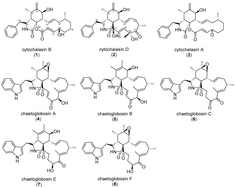
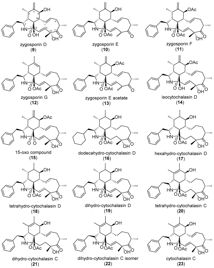
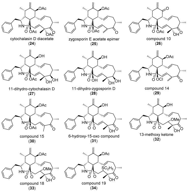
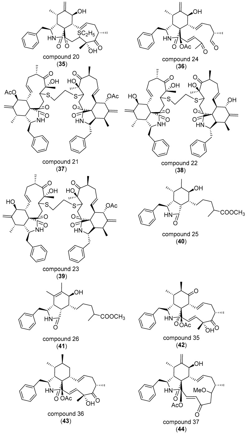
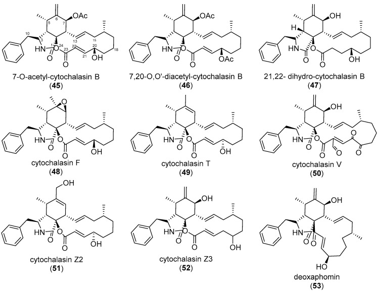
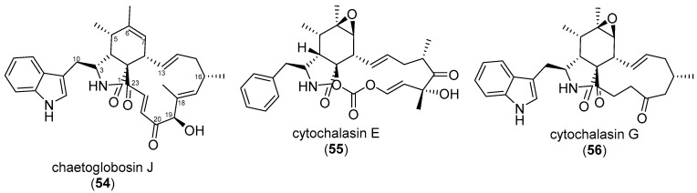
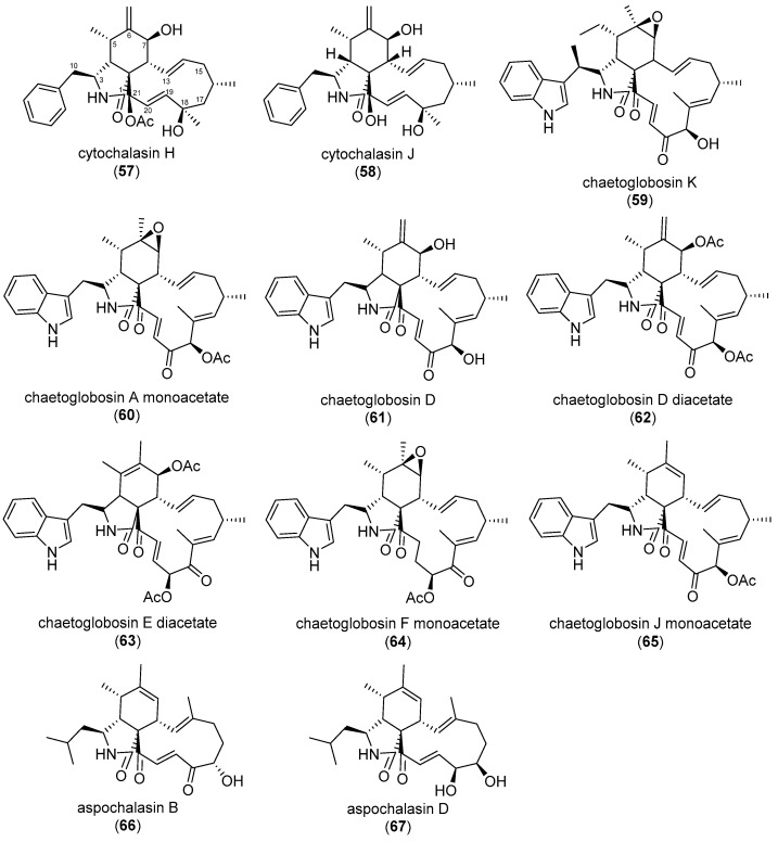
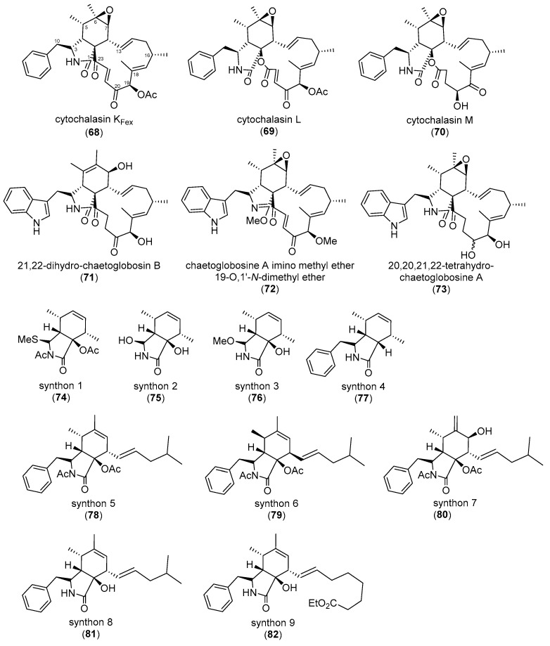
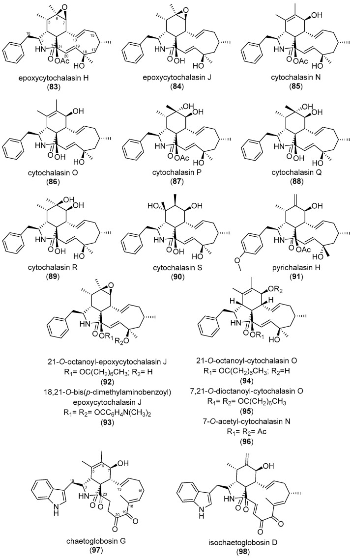
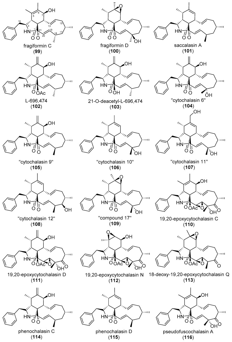
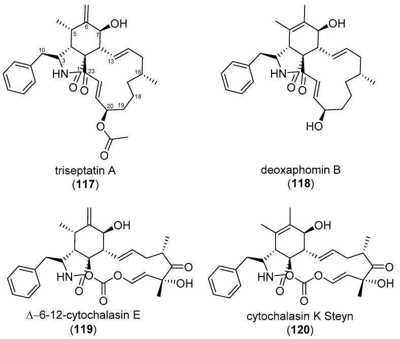
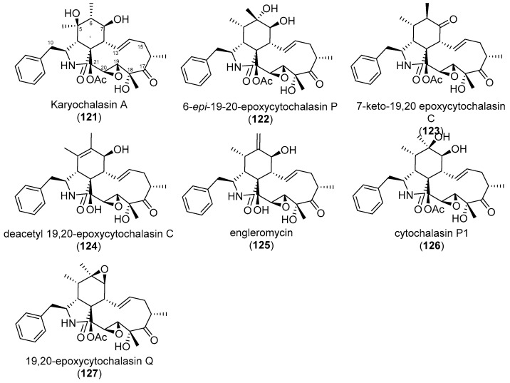
The eukaryotic actin cytoskeleton comprises the protein itself in its monomeric and filamentous forms, G- and F-actin, as well as multiple interaction partners (actin-binding proteins, ABPs). This gives rise to a temporally and spatially controlled, dynamic network, eliciting a plethora of motility-associated processes. To interfere with the complex inter- and intracellular interactions the actin cytoskeleton confers, small molecular inhibitors have been used, foremost of all to study the relevance of actin filaments and their turnover for various cellular processes. The most prominent inhibitors act by, e.g., sequestering monomers or by interfering with the polymerization of new filaments and the elongation of existing filaments. Among these inhibitors used as tool compounds are the cytochalasans, fungal secondary metabolites known for decades and exploited for their F-actin polymerization inhibitory capabilities. In spite of their application as tool compounds for decades, comprehensive data are lacking that explain (i) how the structural deviances of the more than 400 cytochalasans described to date influence their bioactivity mechanistically and (ii) how the intricate network of ABPs reacts (or adapts) to cytochalasan binding. This review thus aims to summarize the information available concerning the structural features of cytochalasans and their influence on the described activities on cell morphology and actin cytoskeleton organization in eukaryotic cells.

Keywords: actin binding proteins; actin inhibitors; chemo-diversity; eukaryotic actin cytoskeleton; secondary metabolites; structure–activity relationship.


Conflict of interest statement

The authors declare no conflict of interest.

Figures

Scheme 1
Scheme 1
Over the past five decades, a multitude of cytochalasin derivatives has been shown to harbor differential efficacy when tested for cytotoxicity and bioactivity in functional cell biological assays towards actin-dependent structures and actin polymerization in biochemical experiments. Modifications at the incorporated amino acid (light-blue box, left), perhydro-isoindolone core (purple box, middle), and macrocyclic ring (light green box, right) have by and large been independently regarded from the remaining cytochalasan backbone (blue, purple, and green lines drawn into a “Vanilla” cytochalasin in the middle top-left). Derivatives featuring a de-aromatized phenylalanine unit were shown to elicit reduced activity (I). Tryptophane-derived indole-substituted compounds were reported to either increase or have no effect on bioactivity (II), while other non-ribosomal peptide substituents have so far not been comprehensively studied (IV). No effect on bioactivity was found for a methyl attached to C-10 (III), or a methoxy group substituted in para position at the phenyl shown in V. Acetylation of a C-7 hydroxy group found in the perhydro-isoindolone core was shown to either decrease or increase efficacy (VI), while substitution with a C-6/C-7 epoxide increased activity (VII). Toxicity was unchanged for a combination of C-7 hydroxy, C-6 methylene, and C-5 methyl moieties (VIII), while a double bond between C-5 and C-6 either had no influence or decreased efficacy compared to the former (IX). Epimers of further hydroxylated compounds were shown to display decreased activity (X). However, compounds lacking oxygen functions in the perhydro-isoindolone core did not differ in efficacy compared to oxygenated ones (XI). The same was reported for a methylated methyl attached to C-11 (XII). Hydroxylation of a keto-group at C-21 was without effect or decreased efficacy (XIII), while acetylation did not change activity, neither when compared to a ketone nor a hydroxy group at C-21 (XIV). Enlargement of the macrocyclic ring by oxygen insertion, forming a lactone, was also without effect (XV). Finally, ring opening and polycyclization were both reported to decrease activity (XVI). For conflicting reports and further structural details, see main text above. Studies cited in the scheme are as follows: Umeda et al. (1975) [55], Minato et al. (1973) [56], Sekita et al. (1985) [57], van Goietsenowen et al. (2011) [58], Yahara et al. (1982) [69], Pourmoghaddham et al. (2022) [78], Hirose et al. (1990) [85], Lambert et al. (2023) [96] and Kemkuignou et al. (2022) [97].
Figure 12
Figure 12
Structures of cytochalasins (128134) analyzed by Kemkuignou et al. (2022) and periconiasin A and E (135136) [97,99,100].
Figure 14
Figure 14
The biotin (“compound 9”, 137), Texas Red (“compound 17”, 138) and Alexa 488 (“compound 19”, 139)-linked pyrichalasin H (91) derivatives generated by click-chemistry following a mutasynthetic approach presented by Wang et al. (2020) [202].
Figure 1
Figure 1
(A). Schematized overview of selected, intracellular actin filament structures and the cellular processes these structures contribute to (filaments and molecules not drawn to scale). The respective positions of filament barbed ends, which cytochalasans are assumed to bind to, are also indicated. Although all actin filament barbed ends are potential targets of cytochalasans, in principle, differential effects on different actin structures can be observed, but the precise molecular reasons for this have yet to be established (for details, see text). (B) Selected, actin-independent activities of cytochalasans. (C). Representative epifluorescence images of human osteosarcoma cells stained for nuclear DNA using DAPI (pseudocolored in blue) and actin filaments with fluorescently-coupled phalloidin (in grey), treated with vehicle control (DMSO, I) or low and high concentrations of CB (1, II + III) for 1 h, with the latter followed by 1 h washout, as indicated (IV). Control U-2 OS cells display discernible stress fibers (I, green arrowhead) and F-actin-rich lamellipodia (I, red arrowhead). Low-dose CB (1) treatments remove lamellipodia at the cell periphery (II, red arrowhead) and cause partial induction of F-actin-rich spots, the precise nature of which is elusive, with stress fibers remaining largely intact (II, green arrowhead). The latter disappear upon high-dose CB (1); aggregates grow even larger (III, orange arrowhead). In spite of these drastic phenotypic changes, a washout for 1 h is sufficient for full recovery of the U-2 OS cell actin network (IV, red arrowhead: lamellipodium, green arrowhead: stress fiber). Figure subpanels (A,B) were created with BioRender.com.
Figure 2
Figure 2
Structures of cytochalasin B (CB, 1), D (CD, 2), A (CA, 3), and chaetoglobosin A-F (ChA-F, 4–8).
Figure 3
Figure 3
Structures of cytochalasin D derivatives (944) tested by Minato et al. (1978) [56].
Figure 3
Figure 3
Structures of cytochalasin D derivatives (944) tested by Minato et al. (1978) [56].
Figure 3
Figure 3
Structures of cytochalasin D derivatives (944) tested by Minato et al. (1978) [56].
Figure 4
Figure 4
Structures of cytochalasans (4553) investigated by van Goietsenoven et al. (2011) [58].
Figure 5
Figure 5
Structures of cytochalasans (5456) tested by Löw et al. (1979) [67].
Figure 6
Figure 6
Structures of cytochalasans (5767) tested by Yahara et al. (1982) [69]. The following compound names were interchangeably used by Sekita et al. (1985): 19-O-acetyl-chaetoglobosin A (60); 7,19-O-diacetyl-chaetoglobosin D (62); 7,19-O-diacetyl-chaetoglobosin E (63); 19-O-acetyl-chaetoglobosin F (64); 19-O-acetyl-chaetoglobosin J (65) [57].
Figure 7
Figure 7
Structures of cytochalasans (6873) and cytochalasin B synthones (7482) analyzed by Sekita et al. (1985) [57].
Figure 8
Figure 8
Structures of cytochalasans (8396) tested by Hirose et al. (1985) [85] and chaetoglobosin G (97) and isochaetoglobosin D (98) by Thohinung et al. 2010 [92].
Figure 9
Figure 9
Structures of cytochalasans tested by Kretz et al. (2010) (99115) and Lambert et al. (2021) (116) [76,78].
Figure 10
Figure 10
Structures of cytochalasans investigated by Garcia et al. (2022) (117, 118) and Pourmoghaddam et al. (2022) (119, 120) [77,79].
Figure 11
Figure 11
Structures of cytochalasans (121127) tested by Lambert et al. (2023) [96].
Figure 13
Figure 13
(A). Number of indexed publications in a PubMed literature search for different members of the cytochalasans. (B). Number of indexed publications in a PubMed literature search for CB (1) vs. CD (2) between January 1967 and June 2023. (C). Number of indexed publications in a PubMed literature search for CA (1), CC (23), CE (55), CF (48), and CH (57) between January 1967 and June 2023. To search for indexed publications for different cytochalasan subclasses, the keywords “alachalasin”, “aspochalasin”, “chaetoglobosin”, “pyrichalasin”, and “trichalasin” were used. For the remainder, “cytochalasin A” was used for CA (1), “cytochalasin B” for CB (2), “cytochalasin C” for CC (23), “cytochalasin F” for CF (48), and “cytochalasin H” for CH (57), spanning a time interval from 1967 until June 2023. Note the overwhelming share of publications indexing either CB (1) or CD (2).

Similar articles

Cited by

References

    1. Macheleidt J., Mattern D.J., Fischer J., Netzker T., Weber J., Schroeckh V., Valiante V., Brakhage A.A. Regulation and role of fungal secondary metabolites. Annu. Rev. Genet. 2016;50:371–392. doi: 10.1146/annurev-genet-120215-035203. - DOI - PubMed
    1. Skellam E. The biosynthesis of cytochalasans. Nat. Prod. Rep. 2017;34:1252–1263. doi: 10.1039/C7NP00036G. - DOI - PubMed
    1. Chepkirui C., Stadler M. The genus Diaporthe: A rich source of diverse and bioactive metabolites. Mycol. Prog. 2017;16:477–494. doi: 10.1007/s11557-017-1288-y. - DOI
    1. Becker K., Stadler M. Recent progress in biodiversity research on the Xylariales and their secondary metabolism. J. Antibiot. 2021;74:1–23. doi: 10.1038/s41429-020-00376-0. - DOI - PMC - PubMed
    1. Charria-Girón E., Surup F., Marin-Felix Y. Diversity of biologically active secondary metabolites in the ascomycete order Sordariales. Mycol. Prog. 2022;21:43. doi: 10.1007/s11557-022-01775-3. - DOI

Publication types

LinkOut - more resources