Review
. 2023 Aug 19;13(16):2670.
doi: 10.3390/ani13162670.

Surgical Anatomy of the Gastrointestinal Tract in Cats

Affiliations
Review

Surgical Anatomy of the Gastrointestinal Tract in Cats

Vasileia Angelou et al. Animals (Basel). .

Abstract

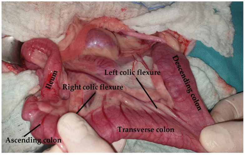
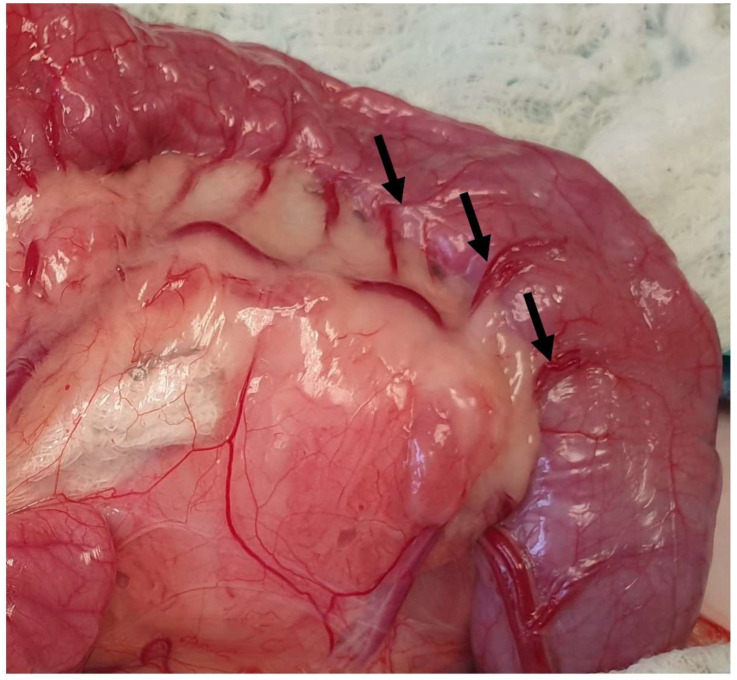
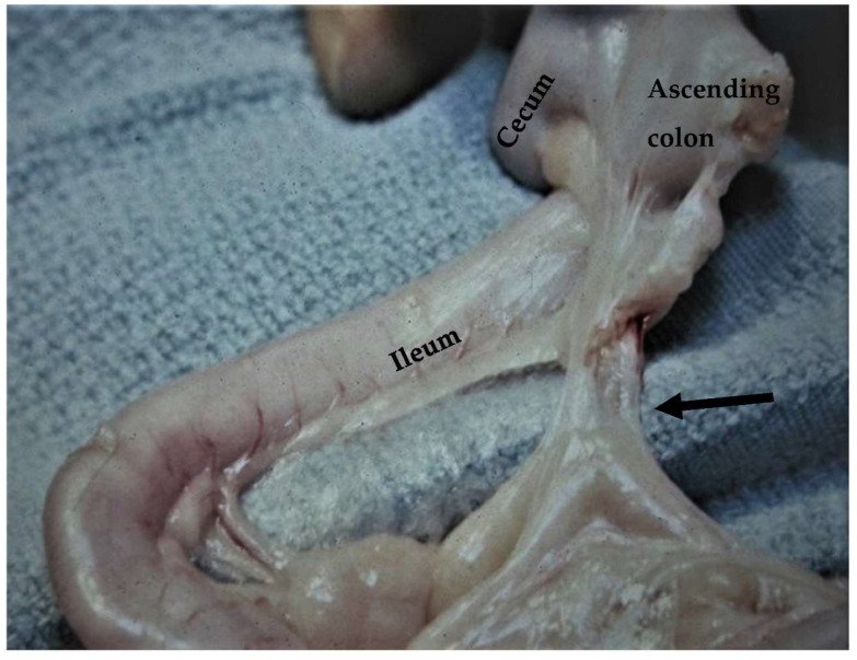
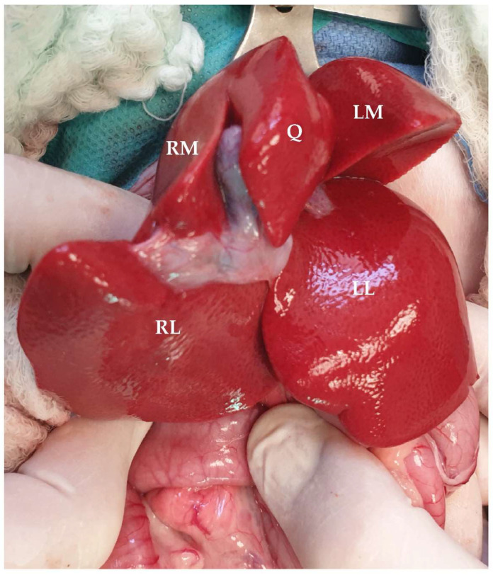
In cats, the gastrointestinal tract is one of the regions in which surgical procedures are most frequently performed by veterinary surgeons; therefore, knowledge of the surgical anatomy of the feline gastrointestinal tract is of high importance. The main surgical procedures performed include gastrotomy, gastrectomy, enterotomy, and enterectomy, as well as procedures in the liver and pancreas. There are also anatomical differences between dogs and cats, increasing the need for deep knowledge of the anatomy treated in the different surgical approaches. The aim of the present review is to describe in detail the anatomy of the gastrointestinal tract in cats highlighting the anatomical regions of significant importance in different surgical procedures.

Keywords: abdomen; anatomy; feline; gastrointestinal surgery.


Conflict of interest statement

The authors declare no conflict of interest.

Figures

Figure 1
Figure 1
The linea alba is identified (arrow) following midline incision in the skin and subcutaneous tissue.
Figure 2
Figure 2
Following midline incision in the ventral abdominal wall, the rectus abdominis muscle (dotted arrow) and the external leaf of the rectus sheath (black arrow) are exposed.
Figure 3
Figure 3
For celiotomy closure, only the external leaf of the rectus sheath should be included in the suture line.
Figure 4
Figure 4
The lesser curvature (arrow), the angular notch (dotted arrow), the greater curvature (short arrow), the pylorus (asterisk), and a gastric lymph node (arrowhead) are visualized. The head is located on the left side of the image.
Figure 5
Figure 5
The left gastric artery (solid arrow) and the right gastric artery (dotted arrow) are exposed following the caudal traction of the stomach using a pair of Babcock forceps. The head is located in the upper part of the image.
Figure 6
Figure 6
The gastroepiploic artery is visualized (arrow) following the ventral traction of the omentum. The head is located on the left side of the image.
Figure 7
Figure 7
The gastric cardia (asterisk) is exposed following caudal traction of the stomach. The esophageal branch of the left gastric artery is evident (arrow). The head is located on the left side of the image.
Figure 8
Figure 8
The gastrotomy is performed in a hypovascular area between the greater and lesser curvatures on the ventral surface of the stomach (dotted line). The head is located on the left side of the image.
Figure 9
Figure 9
Midline celiotomy was performed, and greater (GO) and lesser omentum (LO) were evident. The head is located in the upper part of the image.
Figure 10
Figure 10
The mesoduodenum (asterisk) is retracted to the left to improve visualization of the right sublumbar region (ascending d: ascending duodenum; duodenocolic fold: arrow).
Figure 11
Figure 11
The duodenocolic fold is retracted using Debakey forceps. The head is located on the left side of the image.
Figure 12
Figure 12
The major duodenal papilla is catheterized using a 24-gram venous catheter following a duodenotomy. The head is located on the right side of the image.
Figure 13
Figure 13
The jejunum is exposed. The branches of the cranial mesenteric artery and a jejunal lymph node are evident (asterisk). The vasa recta (arrows) are visualized in a close-up image. The head is located on the left side of the image.
Figure 14
Figure 14
An enterotomy is performed along the antimesenteric region of the jejunum for the removal of a linear foreign body grasped by a hemostat. The head is located on the left side of the image.
Figure 15
Figure 15
The ileum is exposed via traction of the jejunum in a ventral direction. The ileum is identified based on the antimesenteric ileal blood supply (black arrow) and the ileocecal fold (dotted arrow).
Figure 16
Figure 16
The colon is subdivided into ascending, transverse, and descending sections. Major blood supply (black arrow: ileocolic artery; dotted arrow: right colic artery; arrowhead: middle colic artery; pointed arrow: left colic artery; circle pointed arrow: cranial rectal artery) and mesenteric lymph nodes (asterisks) are visualized.
Figure 17
Figure 17
The colon is retracted to the right side of the abdomen to provide access to the left sublumbar region. The head is located on the left side of the image.
Figure 18
Figure 18
Vasa recta of the left colic and caudal mesenteric arteries are exposed (arrows). These vessels may be ligated during a subtotal colectomy.
Figure 19
Figure 19
During subtotal colectomy with preservation of the ileocolic valve, the short mesentery supplying the ileocecocolic region (arrow) may contribute to increased tension at the anastomosis side (courtesy D.M. Bright).
Figure 20
Figure 20
Visceral surface of the feline liver. (RM: right medial lobe; RL: right lateral lobe; Q: quadrate lobe; LM: left medial lobe; LL: left lateral lobe). Head is located in the upper part of the image.
Figure 21
Figure 21
The liver is attached to the diaphragm via the coronary ligaments (pointed arrow). The caudal vena cava (pre-hepatic part) is also visualized (black arrow). The head is located on the left side of the image.
Figure 22
Figure 22
The falciform ligament is lifted using the surgeon’s index finger. The head is located on the left side of the image.
Figure 23
Figure 23
The epiploic foramen (a malleable retractor is inserted into the foramen [white arrow]) is bordered by the caudal vena cava dorsally and the portal vein ventrally. The celiac artery is also visualized. The head is located on the left side of the image.
Figure 24
Figure 24
The omental bursa can be seen by ventrally lifting the greater omentum. The head is located in the upper part of the image.
Figure 25
Figure 25
A hole (black arrows) was created in the ventral leaf of the greater omentum to gain access to the omental bursa. The left pancreatic lobe is lying within the bursa (asterisk). The head is located on the left side of the image.
Figure 26
Figure 26
The portal vein (left branch: dotted arrow; right branch: black arrow) is exposed by ventrally and medially lifting the duodenum.
Figure 27
Figure 27
The gallbladder is exposed by lifting the quadrate lobe of the liver (black arrow: cystic duct; dotted arrow: common bile duct; arrowhead: hepatoduodenal ligament).

Similar articles

Cited by

References

    1. Langley-Hobbs S.J., Demetriou J.L., Ladlow J.F. Preface. In: Langley-Hobbs S.J., Demetriou J., Ladlow J.F., editors. Feline Soft Tissue and General Surgery. Elsevier; London, UK: 2014. p. ix.
    1. Snipes R.L. Anatomy of the cecum of the cat. Anat. Embryol. 1984;170:177–186. doi: 10.1007/BF00319003. - DOI - PubMed
    1. Eken E., Gezici M. The Influence of Stomach Volume on the Liver Topography in Cats. Anat. Histol. Embryol. 2002;31:99–104. doi: 10.1046/j.1439-0264.2002.00366.x. - DOI - PubMed
    1. Hayes G. Gastrointestinal foreign bodies in dogs and cats: A retrospective study of 208 cases. J. Small Anim. Pract. 2009;50:576–583. doi: 10.1111/j.1748-5827.2009.00783.x. - DOI - PubMed
    1. Cariou M.P., Halfacree Z.J., Lee K.C., Baines S.J. Successful surgical management of spontaneous gastric perforations in three cats. J. Feline Med. Surg. 2010;12:36–41. doi: 10.1016/j.jfms.2009.12.005. - DOI - PMC - PubMed

LinkOut - more resources