Review
. 2023 Jul 28;12(8):1517.
doi: 10.3390/antiox12081517.

Mitochondrial Volume Regulation and Swelling Mechanisms in Cardiomyocytes

Affiliations
Review

Mitochondrial Volume Regulation and Swelling Mechanisms in Cardiomyocytes

Xavier R Chapa-Dubocq et al. Antioxidants (Basel). .

Abstract

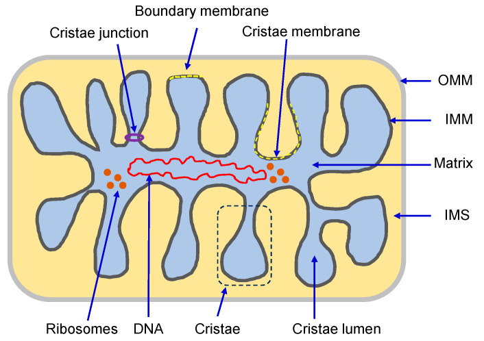
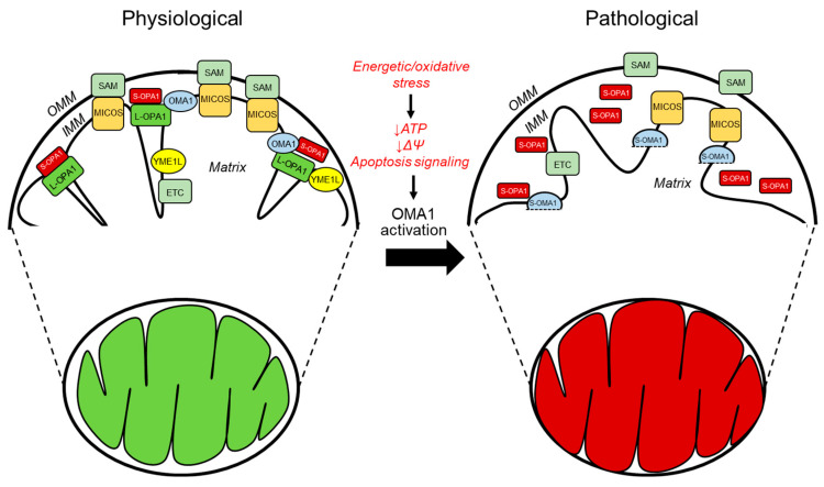
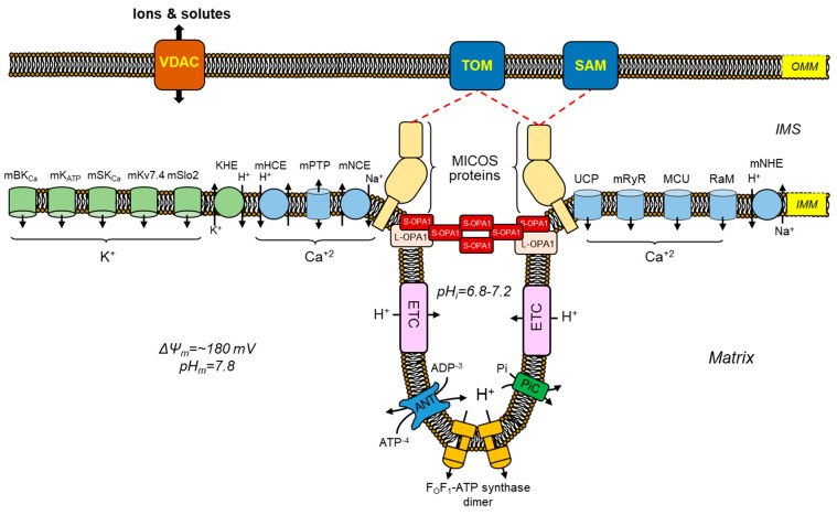
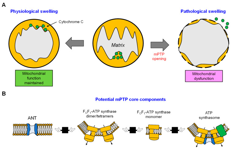
Mitochondrion, known as the "powerhouse" of the cell, regulates ion homeostasis, redox state, cell proliferation and differentiation, and lipid synthesis. The inner mitochondrial membrane (IMM) controls mitochondrial metabolism and function. It possesses high levels of proteins that account for ~70% of the membrane mass and are involved in the electron transport chain, oxidative phosphorylation, energy transfer, and ion transport, among others. The mitochondrial matrix volume plays a crucial role in IMM remodeling. Several ion transport mechanisms, particularly K+ and Ca2+, regulate matrix volume. Small increases in matrix volume through IMM alterations can activate mitochondrial respiration, whereas excessive swelling can impair the IMM topology and initiates mitochondria-mediated cell death. The opening of mitochondrial permeability transition pores, the well-characterized phenomenon with unknown molecular identity, in low- and high-conductance modes are involved in physiological and pathological increases of matrix volume. Despite extensive studies, the precise mechanisms underlying changes in matrix volume and IMM structural remodeling in response to energy and oxidative stressors remain unknown. This review summarizes and discusses previous studies on the mechanisms involved in regulating mitochondrial matrix volume, IMM remodeling, and the crosstalk between these processes.

Keywords: heart; ions; ischemia-reperfusion; mitochondria; permeability transition pore.


Conflict of interest statement

The authors declare no conflict of interest.

Figures

Figure 1
Figure 1
Structure of mitochondria (see text for details).
Figure 2
Figure 2
The primary regulators of mitochondrial cristae organization in both physiological and pathological conditions. The diagram depicts the structural arrangement of mitochondrial membranes, in which the cristae arise from the inward folding of the inner membrane towards the matrix. The MICOS complex, which is responsible for organizing mitochondrial contact sites and cristae, is situated at the CJs. It comprises seven subunits, namely, MIC10, MIC13, MIC19, MIC25, MIC26, MIC27, and MIC60. The stabilization of the CJs and establishing contacts between the IMM and OMM requires MICOS to be involved. The convergence of MICOS and the SAM complex results in the formation of a more extensive entity known as the mitochondrial intermembrane space bridging complex, which encompasses the intermembrane space. The protein OPA1 is highly localized at the CJs and its presence is crucial for preserving the proper dimensions of these structures. This necessitates the interplay between the membrane-bound long (L-) variants and the soluble short (S-) variants of OPA1. Under physiological (denoted in green) and pathological (denoted in red) conditions, the IMM proteases YME1L and OMA1 regulate OPA1. The protein OMA1 is responsible for cleaving all isoforms of OPA1 at the S1 site, whereas YME1L is responsible for cleaving OPA1 at the S2 site but only for the subset of OPA1 that contains the splice variant. Under physiological conditions, YME1L modifies the L-OPA1 to S-OPA1 ratio of the heterooligomeric complexes by cleaving a subset of the L-OPA1 isoforms at the S2 cleavage site. Under normal circumstances, OMA1 exhibits minimal activity; however, it is known to become activated in response to pathological conditions. OMA1 cleaves all L-OPA1 isoforms at the S1 cleavage site, releasing S-OPA1 from the IMM and OMA1 self-cleavage resulting in S-OMA1. Additionally, under stress conditions, the SAM-Mic19-Mic-60 axis can be disrupted through the cleavage of Mic19 by OMA1, resulting in the separation of the SAM and MICOS. Overall, the activation of OMA1 under pathological circumstances leads to impaired mitochondrial function.
Figure 3
Figure 3
Mechanisms involved in maintaining ion homeostasis, structural maintenance, and matrix volume within the mitochondria. A graphical representation that illustrates the primary influx and efflux channels of Ca2+ and K+, which are responsible for regulating the volume, as well as the structural constituents that govern the morphology of cardiac mitochondria. The primary mechanisms responsible for Ca2+ influx are the MCU, RaM, mRyR, and uncoupling proteins 2 and 3 (UCP2/3). The maintenance of Ca2+ and ion homeostasis in the matrix is a crucial aspect, wherein the Ca2+ efflux mechanisms such as mitochondrial mNCE, mHCE, and mPTP play a significant role. The transportation of K+ holds equal significance for the metabolism and functioning of mitochondria. Alterations in the concentration of K+ exhibit a direct correlation with variations in the volume of the mitochondrial matrix. The mechanisms responsible for K+ influx comprise the mBKCa, mKATP, mSKCa, mKv7.4, and mSlo2. Conversely, the K+ efflux mechanisms are restricted to the KHE. The function of OPA1 is the fusion of IMM and its regulatory role in cristae morphogenesis. In the heart, five isoforms of OPA1 exist that can be characterized as either L-OPA1 to S-OPA1. The various isoforms of OPA1 have demonstrated the capacity to oligomerize and uphold stringent CJs. A protein complex known as MICOS facilitates preserving a stable state in mitochondrial cristae junctions. In addition to its membrane bending capabilities, MICOS exhibits interactions with proteins situated in the OMM, including the translocase of the outer membrane (TOM) complex, the sorting and assembly machinery (SAM), and the voltage-dependent anion channel (VDAC) protein. Furthermore, the mitochondrial Na+/H+ exchanger (mNHE) has been implicated in the maintenance of ion homeostasis within mitochondria. Also, the invaginations of CJs are generated by dimeric FOF1-ATP synthase, where CJ structures serve as the site for ETC complexes, ANT, and phosphate carrier (PiC), which utilize the pH gradient to facilitate the process of ATP synthesis.
Figure 4
Figure 4
Publication history related to mitochondria and mPTP research. (A) Mitochondrial research conducted over time, indicating a steady increase in the number of studies. (B) A decline of approximately eight years in mPTP research and mPTP-derived therapeutic interventions. The data was obtained from PubMed (https://www.ncbi.nlm.nih.gov (accessed on 5 June 2023)).
Figure 5
Figure 5
mPTP-mediated matrix swelling and potential mPTP models. (A) The opening of the mitochondrial permeability transition pore (mPTP) triggers mitochondrial swelling, leading to various consequences for mitochondrial function and cell survival, depending on the degree of swelling. (B) Although the molecular identification of the mPTP remains unclear, it is widely accepted that CypD acts as a protein regulator of the mPTP. Most studies have focused on a pair of proteins known as ANT and FOF1-ATP synthase (F-ATP synthase). The formation of the mPTP may be attributed to the synergistic or independent functioning of ANT and FOF1-ATP synthase. However, the participation of both entities is essential. It can be proposed that mPTP may be formed by one of the proteins or complexes, namely, ANT, FOF1-ATP synthase, FOF1-ATP synthase dimer/tetramer, or the ATP synthasome.

Similar articles

Cited by

References

    1. Ingwall J.S. Is creatine kinase a target for AMP-activated protein kinase in the heart? J. Mol. Cell Cardiol. 2002;34:1111–1120. doi: 10.1006/jmcc.2002.2062. - DOI - PubMed
    1. Varikmaa M., Guzun R., Grichine A., Gonzalez-Granillo M., Usson Y., Boucher F., Kaambre T., Saks V. Matters of the heart in bioenergetics: Mitochondrial fusion into continuous reticulum is not needed for maximal respiratory activity. J. Bioenerg. Biomembr. 2013;45:319–331. doi: 10.1007/s10863-012-9494-4. - DOI - PubMed
    1. Kuznetsov A.V., Javadov S., Margreiter R., Grimm M., Hagenbuchner J., Ausserlechner M.J. The Role of Mitochondria in the Mechanisms of Cardiac Ischemia-Reperfusion Injury. Antioxidants. 2019;8:454. doi: 10.3390/antiox8100454. - DOI - PMC - PubMed
    1. Murphy E., Ardehali H., Balaban R.S., DiLisa F., Dorn G.W., 2nd, Kitsis R.N., Otsu K., Ping P., Rizzuto R., Sack M.N., et al. Mitochondrial Function, Biology, and Role in Disease: A Scientific Statement From the American Heart Association. Circ. Res. 2016;118:1960–1991. doi: 10.1161/RES.0000000000000104. - DOI - PMC - PubMed
    1. Gustafsson A.B., Gottlieb R.A. Heart mitochondria: Gates of life and death. Cardiovasc. Res. 2008;77:334–343. doi: 10.1093/cvr/cvm005. - DOI - PubMed

LinkOut - more resources