Review
. 2023 Aug 21;13(16):2715.
doi: 10.3390/diagnostics13162715.

Keratoconus Diagnosis: From Fundamentals to Artificial Intelligence: A Systematic Narrative Review

Affiliations
Review

Keratoconus Diagnosis: From Fundamentals to Artificial Intelligence: A Systematic Narrative Review

Sana Niazi et al. Diagnostics (Basel). .

Abstract

The remarkable recent advances in managing keratoconus, the most common corneal ectasia, encouraged researchers to conduct further studies on the disease. Despite the abundance of information about keratoconus, debates persist regarding the detection of mild cases. Early detection plays a crucial role in facilitating less invasive treatments. This review encompasses corneal data ranging from the basic sciences to the application of artificial intelligence in keratoconus patients. Diagnostic systems utilize automated decision trees, support vector machines, and various types of neural networks, incorporating input from various corneal imaging equipment. Although the integration of artificial intelligence techniques into corneal imaging devices may take time, their popularity in clinical practice is increasing. Most of the studies reviewed herein demonstrate a high discriminatory power between normal and keratoconus cases, with a relatively lower discriminatory power for subclinical keratoconus.

Keywords: artificial intelligence; biomechanical phenomena; computer; corneal topography; deep learning; diagnosis; keratoconus; machine learning; neural networks; optical coherence.


Conflict of interest statement

No conflicting relationship exists for any author. All authors completed and submitted the ICMJE disclosure of interest form.

Figures

Figure 1
Figure 1
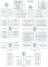
A structured overview of articles about keratoconus and artificial intelligence. Topographic Modeling System (TMS); Optical Path Difference (OPD) Scan; Ultra-High Resolution Optical Coherence Tomography (UHR-OCT) [2,10,11,21,22,23,24,25,26,27,28,29,30,31,32,33,34,35,36,37,38,39,40,41].
Figure 2
Figure 2
A comparison of the broad spectrum of artificial datasets about sensitivity, specificity, and accuracy represented by different machines in keratoconus. Biomechanically Corrected Intraocular Pressure (bIOP); First and Second Applanation (A1, A2); Time (T); Velocity (V); Deformation Amplitude (DA); Deflection Length (DL); Deflection Amplitude (DeflA); Delta Arc Length (dArclength); Vinciguerra Screening Parameters (VSP); Ambrósio’s Relational Thickness to the Horizontal Profile (ARTh); Integrated Radius (INR/IR); Stiffness Parameter (SP); Corvis Biomechanical Index (CBI); Radius (Rad); Highest Concavity (HC); Peak Distance (PD); Index of Height Asymmetry (IHA); Index of Surface Variance (ISV); Maximum Keratometry front (Kmax); Difference in Maximum–Minimum Anterior Elevation above/below the Best-Fit Sphere (Aedif); Index of Height Decentration (IHD); Minimal Sagittal Curvature (Rmin); Index of Vertical Asymmetry (IVA); Posterior Elevation (PE); Minimum Keratometry (Kmin); Maximum Posterior Elevation in 5 mm Zone above the Best-Fit Sphere (PE); Difference in Maximum–Minimum Posterior Elevation above/below the Best-Fit Sphere (Pedif); Maximum Anterior Elevation in 5 mm Zone above the Best-Fit Sphere (AE); Thickness at the Corneal Apex (AT); Corneal Thickness at the Pupil Center (PT); Minimum Sagittal Curvature (Rmin); Minimum Pachymetric Progression Index (RPImin); Average Pachymetric Progression Index (RPIavg); Maximum Pachymetric Progression Index (RPImax); Anterior Chamber Depth (ACD); Corneal Volume (CV); Maximum Ambrósio Relational Thickness (ARTmax); Average Ambrósio Relational Thickness (ARTavg); Minimum Corneal Thickness (MT); Central KC Index (CKI); Anterior Chamber Volume (ACV); Belin–Ambrósio Enhanced Ectasia Total Deviation value (BAD_D); Surface Regularity Index (SRI); Standard Deviation of Corneal Power (SDP); Opposite Sector Index (OSI); Surface Asymmetry Index (SAI); Percentage Probability of KC (PPK); KC Prediction Index (KPI); Asphericity Asymmetry Index (AAI); Differential Sector Index (DSI); Inferior–Superior (I–S) Index; Total Corneal Power (TCP); Center/Surround Index (CSI); Irregular Astigmatism Index (IAI); Root Mean Square (RMS); Baiocchi Calossi Versaci (BCV); Posterior Corneal Aberrations (BCVb); Thinnest Point of the Cornea (ThkMin); Total Wavefront Error (TWFE); Spherical Aberrations (SA); KC Vertex back (KVb); Higher Order Aberration (HOA); Thinnest Corneal Thickness (TCT); Machine Learning (ML); Multilayer Perceptron (MLP); Screening Corneal Objective Risk of Ectasia (SCORE); Support Vector Machine (SVM); Logistic Regression (LR); Fourier-Incorporated KC Detection Index (FKI); Standard Deviation of Thickness Profile between Individual and Normal Patterns of Epithelium, Bowman’s Layer, and Stroma (EPSD, BPSD, SPSD); Profile Variation in epithelium, Bowman’s Layer, or Stroma Thickness Profile within Each Individual (EPV, BPV, SPV); Ectasia Index of Epithelium, Bowman’s Layer, or Stroma (EEI, BEI, SEI); Maximum Ectasia Index of Epithelium Layer, Bowman’s Layer, or Stroma (EEI-MAX, BEI-MAX, SEI-MAX); Mean Thickness of Epithelium, Bowman’s Layer, or Stroma (EMean, BMean, SMean); Thinnest Thickness of the Inferior Epithelium, Bowman’s Layer, or Stroma Thickness Map (Emin, Bmin, Smin); Thickest Thickness of the Superior Epithelium, Bowman’s Layer, or Stroma Thickness Map (Emax, Bmax, Smax).
Figure 3
Figure 3
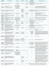
Abnormal and suggestive of KC thresholds for different devices. Best-Fit Sphere (BFS); Asphericity Asymmetry Index (AAI); Center/Surround Index (CSI); Differential Sector Index (DSI); Irregular Astigmatism Index (IAI); Inferior–Superior (I–S) Index; KC Prediction Index (KPI); Opposite Sector Index (OSI); Percentage Probability of KC (PPK); Standard Deviation of Corneal Power (SDP); Surface Asymmetry Index (SAI), Surface Regularity Index (SRI); Total Corneal Power (TCP); Central KC Index (CKI); KC Index (KI); Index of Height Asymmetry (IHA); Index of Height Decentration (IHD); Pentacam Topographical KC Classification (TKC); Index of Surface Variance (ISV); Index of Vertical Asymmetry (IVA); Minimal Sagittal Curvature (Rmin); Posterior Elevation (PE); Ambrósio’s Relational Thickness (ART); Belin–Ambrósio Enhanced Ectasia Display Total Deviation (BAD_D) Value; Central Corneal Thickness (CCT); Pachymetric Progression Indices (PPI); Thinnest Corneal Thickness (TCT); Maximum Keratometry (Kmax); Relative Skewing of the Steepest Radial Axes (SRAX); Epithelium Profile Variation (EPV); Bowman’s Layer Profile Variation (BPV); Maximum Elevation (Emax); Central Elevation (Ecenter); Epithelium Profile Standard Deviation (EPSD); Maximum Ectasia Index of Bowman’s Layer (BEI-MAX); Thinnest Thickness of the Inferior Bowman’s Layer Thickness Map (Bmin); Best-Fit Sphere (BFS). * and **: Although several studies have discussed these parameters, there is no consequence on the thresholds and cutoff values.
Figure 4
Figure 4
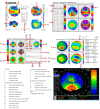
The important feature of four devices in a keratoconus eye from one patient. The Orbscan II, the Pentacam AXL/wave (four maps refractive display), Belin/Ambrosio enhanced ectasia display with a pachymetric map, the Galilei G4, and axial map Sirius the lowest part).
Figure 5
Figure 5
Comparison of epithelial thickness measurements, RTVue SD-OCT device (Optovue, Inc., Fremont, CA, USA), and epithelial thickness map of a patient after Smile’s derived lenticule implantation.
Figure 6
Figure 6
Factors affecting corneal biomechanical properties. Laser-Assisted In Situ Keratomileusis (LASIK); Epithelial LASIK (Epi-LASIK); Small Incision Lenticule Extraction (SMILE); IntraCorneal Ring Segments (ICRS); Corneal Collagen Crosslinking (CXL); Phakic Intraocular Lens (pIOL); Central Corneal Thickness (CCT); Intraocular Pressure (IOP).
Figure 7
Figure 7
Study selection according to search strategy.
Figure 8
Figure 8
Summary of automatic screening, diagnosis, and classification methods for KC. Patients (p); Eyes (e); Group (g); KC group (KCG); control group (CG); Normal (Nl); Follow up (F/U); Keratoconus (KC); Clinical KC (CKC); Subclinical KC (SKC); Advanced KC (AKC); very asymmetric ectasia (VAE); Very Asymmetric Ectasia but with Normal Corneal Topography (VAE-NTG); Ocular Surface Disorders (OSD); Epithelial Basement Membrane Dystrophy (EBMD); Dry Eye Disease (DED); Keratitis Precipitate (KP), Subepithelial Opacity (SEO); Area Under the Curve (AUC); Area Under the Receiver Operator Characteristic Curve (AUROCC); Accuracy (AC); Sensitivity (Sen); Specificity (Spe); Recall (R); Purity (Pu); Belin–Ambrósio Deviation Index (BAD-D); Corneal Tomography Multivariate Index (CTMVI); Pentacam Topographical KC Classification (TKC); Tomographic–Biomechanical Parameter (TBI); Zernike Coefficients (ZC); Corneal Epithelial Thickness (ET); Deformation Amplitude (DA); Peak Distance (PD) at the Highest Concavity; Boosted Ectasia Susceptibility Tomography Index (BESTi); Multiple Logistic Regression Analysis (MLRA); Artificial intelligence (AI); Paraconsistent Feature Engineering (PFE); Support Vector Machine (SVM); Pentacam Random Forest index (PRFI); Artificial Neural Network (ANN); Flower Pollination Algorithm (FPA); Random Forest (RF); Radial Basis Function NN (RBFNN); Particle Swarm Optimization (PSO); Fractional Order PSO (FPSO); Discrete PSO (DPSO); Linear Regression (LR); Time Delay Neural Network (TDNN); Convolutional NN (CNN); Feedforward Neural Network (FNN); Multilayer Perceptron (MLP); Local Binary Pattern (LBP); Local Directional Pattern (LDP); Local Optimal Oriented Pattern (LOOP); Cat Swarm Optimization (CSO); Linear Discriminant Analysis (LDA); Principal Component Analysis (PCA); Quadratic Discriminant Analysis (QDA); Masked Face Analysis (MAFA); Anterior Segment Optical Coherence Tomography (AS-OCT); Photorefractive Keratectomy (PRK); Phototherapeutic Keratectomy (PTK); Penetrating Keratoplasty (PK); Lamellar Keratoplasty (LK); Laser-Assisted In Situ Keratomileusis (LASIK); Post-LASIK Ectasia (PLE). [17,20,25,26,27,28,29,32,37,41,47,48,49,56,75,86,91,95,118,119,130,131,132,137,138,139,140,141,142,143,144,145,146,147,148,149].
Figure 8
Figure 8
Summary of automatic screening, diagnosis, and classification methods for KC. Patients (p); Eyes (e); Group (g); KC group (KCG); control group (CG); Normal (Nl); Follow up (F/U); Keratoconus (KC); Clinical KC (CKC); Subclinical KC (SKC); Advanced KC (AKC); very asymmetric ectasia (VAE); Very Asymmetric Ectasia but with Normal Corneal Topography (VAE-NTG); Ocular Surface Disorders (OSD); Epithelial Basement Membrane Dystrophy (EBMD); Dry Eye Disease (DED); Keratitis Precipitate (KP), Subepithelial Opacity (SEO); Area Under the Curve (AUC); Area Under the Receiver Operator Characteristic Curve (AUROCC); Accuracy (AC); Sensitivity (Sen); Specificity (Spe); Recall (R); Purity (Pu); Belin–Ambrósio Deviation Index (BAD-D); Corneal Tomography Multivariate Index (CTMVI); Pentacam Topographical KC Classification (TKC); Tomographic–Biomechanical Parameter (TBI); Zernike Coefficients (ZC); Corneal Epithelial Thickness (ET); Deformation Amplitude (DA); Peak Distance (PD) at the Highest Concavity; Boosted Ectasia Susceptibility Tomography Index (BESTi); Multiple Logistic Regression Analysis (MLRA); Artificial intelligence (AI); Paraconsistent Feature Engineering (PFE); Support Vector Machine (SVM); Pentacam Random Forest index (PRFI); Artificial Neural Network (ANN); Flower Pollination Algorithm (FPA); Random Forest (RF); Radial Basis Function NN (RBFNN); Particle Swarm Optimization (PSO); Fractional Order PSO (FPSO); Discrete PSO (DPSO); Linear Regression (LR); Time Delay Neural Network (TDNN); Convolutional NN (CNN); Feedforward Neural Network (FNN); Multilayer Perceptron (MLP); Local Binary Pattern (LBP); Local Directional Pattern (LDP); Local Optimal Oriented Pattern (LOOP); Cat Swarm Optimization (CSO); Linear Discriminant Analysis (LDA); Principal Component Analysis (PCA); Quadratic Discriminant Analysis (QDA); Masked Face Analysis (MAFA); Anterior Segment Optical Coherence Tomography (AS-OCT); Photorefractive Keratectomy (PRK); Phototherapeutic Keratectomy (PTK); Penetrating Keratoplasty (PK); Lamellar Keratoplasty (LK); Laser-Assisted In Situ Keratomileusis (LASIK); Post-LASIK Ectasia (PLE). [17,20,25,26,27,28,29,32,37,41,47,48,49,56,75,86,91,95,118,119,130,131,132,137,138,139,140,141,142,143,144,145,146,147,148,149].

Similar articles

Cited by

References

    1. Whitcher J.P., Srinivasan M., Upadhyay M.P. Corneal blindness: A global perspective. Bull. World Health Organ. 2001;79:214–221. - PMC - PubMed
    1. Cao K., Verspoor K., Sahebjada S., Baird P.N. Accuracy of Machine Learning Assisted Detection of Keratoconus: A Systematic Review and Meta-Analysis. J. Clin. Med. 2022;11:478. doi: 10.3390/jcm11030478. - DOI - PMC - PubMed
    1. Gatinel D., Galvis V., Tello A., Niño C.A., Rey J.J., Camacho P.A., Serrano S.E. Obstructive Sleep Apnea–Hypopnea Syndrome and Keratoconus: An Epiphenomenon Related to Sleep Position? Cornea. 2020;39:e11–e12. doi: 10.1097/ICO.0000000000002219. - DOI - PubMed
    1. Gurnani B., Kaur K. Evolving concepts in etiopathogenesis of keratoconus: Is it quasi-inflammatory or inflammatory? Indian J. Ophthalmol. 2023;71:2609–2610. doi: 10.4103/ijo.IJO_783_22. - DOI - PMC - PubMed
    1. Seth I., Bulloch G., Vine M., Outmezguine J., Seth N., Every J., Daniell M. The association between keratoconus and allergic eye diseases: A systematic review and meta-analysis. Clin. Exp. Ophthalmol. 2023;51:O1–O16. doi: 10.1111/ceo.14215. - DOI - PubMed

LinkOut - more resources