. 2023 Aug 18;12(16):3109.
doi: 10.3390/foods12163109.

The Protective Effect and Mechanism of a Phytochemical Extract from the Wild Vegetable Shutou (Crateva unilocularis Buch.) against Acetaminophen-Induced Liver Injury in Mice

Affiliations

The Protective Effect and Mechanism of a Phytochemical Extract from the Wild Vegetable Shutou (Crateva unilocularis Buch.) against Acetaminophen-Induced Liver Injury in Mice

Meimei Shan et al. Foods. .

Abstract

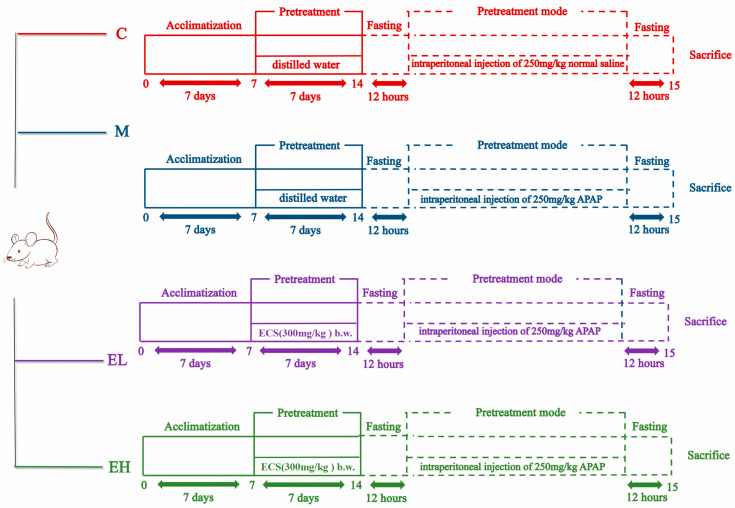
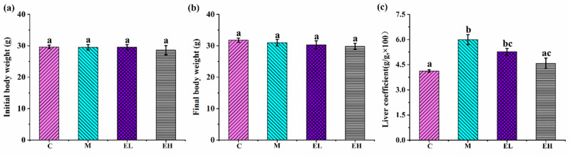
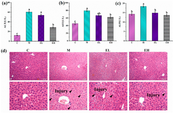
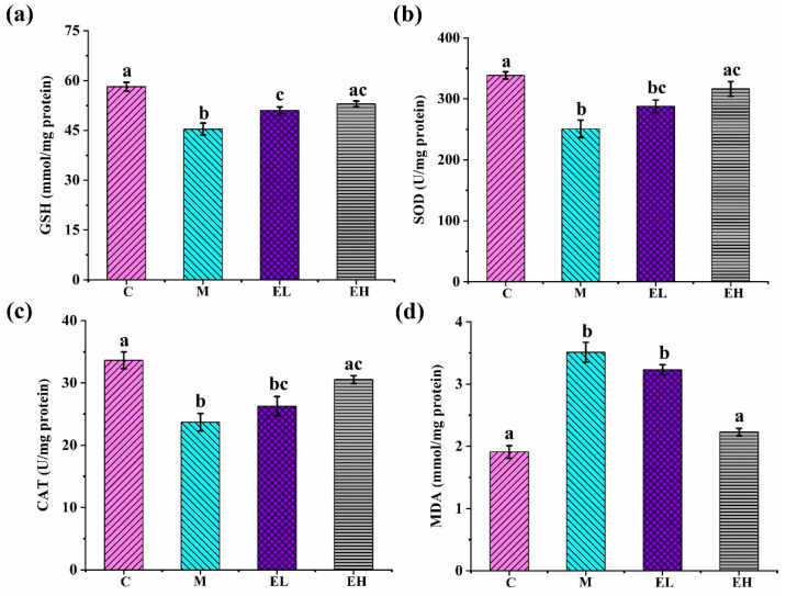
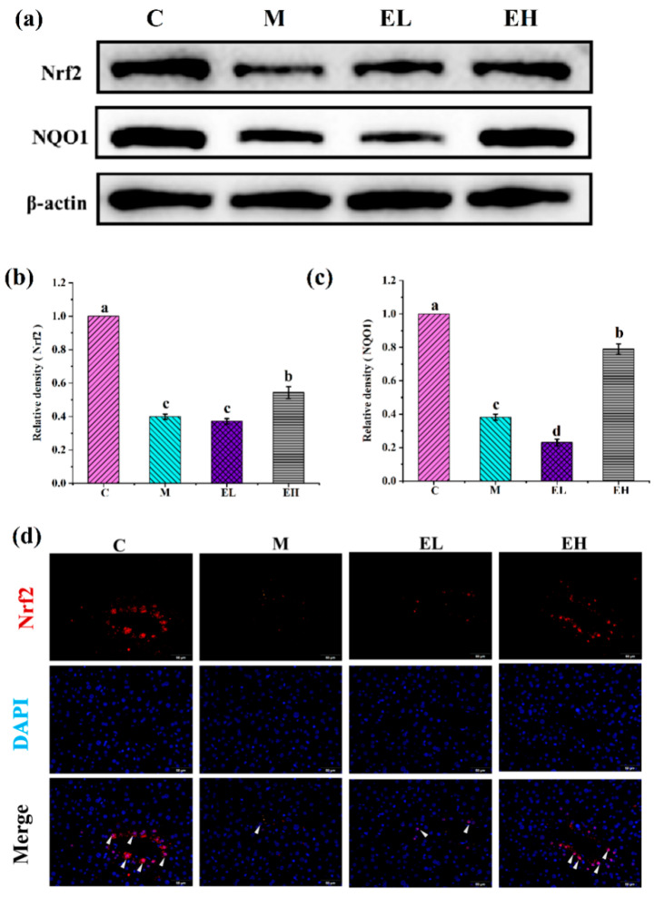
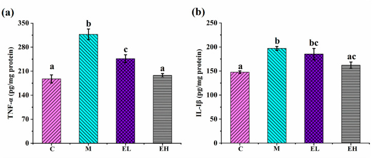
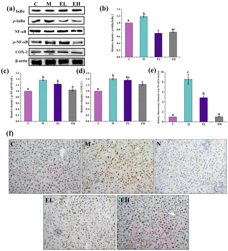
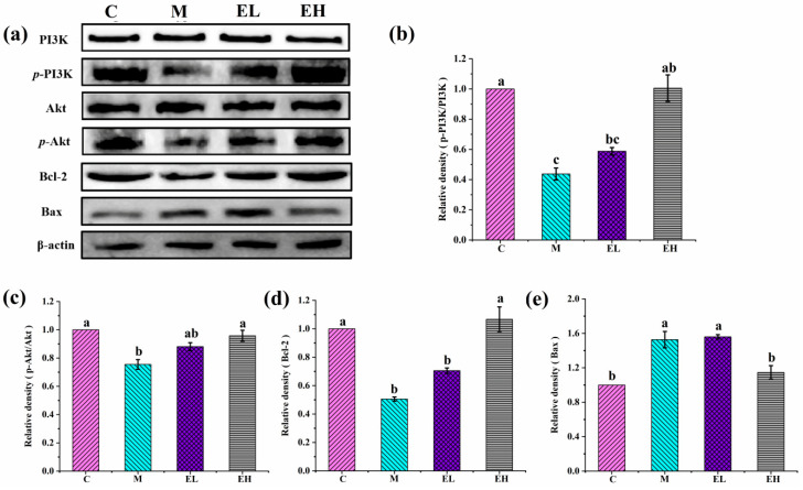
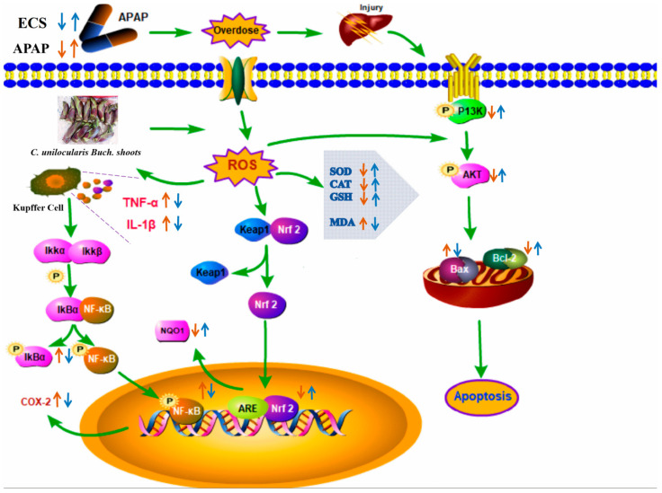
Acetaminophen (APAP) abuse is a common public health problem which can cause severe liver damage. However, strategies for dealing with this situation safely and effectively are very limited. The goal of the current work was to evaluate the protection and potential molecular mechanisms of an ethanol extract from shoots of the wild vegetable shutou (Crateva unilocularis Buch.) (ECS) against APAP-induced liver damage in mice. Mice orally received ECS for seven days (300 or 600 mg/kg b.w. per day) before being intraperitoneally injected with APAP (250 mg/kg). Results exhibited that ECS obviously decreased the content of alkaline phosphatase, alanine aminotransferase, aspartate transaminase, and malondialdehyde (p < 0.05). Catalase and superoxide dismutase were notably restored (p < 0.05), and the content of reduced glutathione was obviously increased (p < 0.05). Moreover, ECS significantly inhibited the secretion of interleukin-1β and tumor necrosis factor-α (p < 0.05). Further analyses of the mechanisms showed that ECS may alleviate oxidative stress in the liver by increasing the expression of the nuclear factor erythroid-2-related factor 2 and NADH quinone oxidoreductase 1 proteins, and may suppress liver inflammation by inhibiting the expression of the phosphorylated-inhibitor kappa B alpha/inhibitor kappa B alpha, phosphorylated-nuclear factor κB/nuclear factor κB, and cyclooxygenase-2 proteins. Meanwhile, ECS inhibited hepatocyte apoptosis by enhancing B-cell lymphoma gene 2 and suppressing Bcl-2-associated X protein. In summary, ECS may be used as a dietary supplement to prevent the liver damage caused by APAP abuse.

Keywords: APAP; Crateva unilocularis Buch.; liver damage; reactive oxygen species.


Conflict of interest statement

The authors declare that they have no known competing financial interests or personal relationships that could have appeared to influence the work reported in this paper.

Figures

Figure 1
Figure 1
Outline of the experiment on the effect of ECS on ALI caused by APAP. C, M, EL, and EH represent the control, model, low-dose, and high-dose ECS groups (300 or 600 mg kg−1 of b.w.), respectively.
Figure 2
Figure 2
Influence of ECS on the body weight and liver coefficient in mice with APAP-induced ALI: (a,b) initial and final body weight, respectively, and (c) liver coefficient. Data are presented as the mean ± S.E. (n = 10). For each biochemical indicator, different letters suggest significant differences (p < 0.05). C, M, EL, and EH represent control, model, low-dose, and high-dose ECS groups (300 or 600 mg kg−1 of b.w.), respectively.
Figure 3
Figure 3
Effects of ECS on (a) ALT, (b) AST, and (c) ALP in the plasma along with (d) liver histopathological changes as observed by H&E staining. Data are presented as the mean ± S.E. (n = 10). For each biochemical indicator, different letters suggest significant differences (p < 0.05). ALP, ALT, and AST represent alkaline phosphatase, alanine, aminotransferase, and aspartate transaminase, respectively, while C, M, EL, and EH represent the control, model, low-dose, and high-dose ECS groups (300 or 600 mg kg−1 of b.w.), respectively.
Figure 4
Figure 4
The effects of ECS on (a) GSH, (b) SOD, (c) CAT, and (d) MDA in the liver tissue. Data are presented as the mean ± S.E. (n = 10). For each biochemical indicator, different letters suggest significant differences (p < 0.05). SOD, superoxide dismutase; GSH, reduced glutathione; CAT, catalase; MDA, malondialdehyde; C, M, EL, and EH represent control, model, low-dose, and high-dose ECS groups (300 or 600 mg kg−1 of b.w.), respectively.
Figure 5
Figure 5
Effects of ECS on the Nrf2 and NQO1 proteins and immunofluorescence of the nuclear transfer of Nrf2 in the livers of mice with or without APAP treatment. (a) Immunoblotting analysis of the Nrf2 and NQO1 proteins. The relative contents of (b) Nrf2 and (c) NQO1 normalized with β-actin and Group C, expressed by the grayscale. (d) The Nrf2 immunofluorescence results entering the nucleus. Data are presented as the mean ± S.E. (n = 4). For each protein, different letters suggest significant differences (p < 0.05). Nrf2 and NQO1 represent nuclear factor erythroid-2 related factor 2 and NADH quinone oxidoreductase 1, respectively; C, M, EL, and EH represent control, model, low-dose, and high-dose ECS groups (300 or 600 mg kg−1 of b.w.), respectively.
Figure 6
Figure 6
Effects of ECS on (a) TNF-α and (b) IL-1β in the liver tissue. Data are presented as the mean ± S.E. (n = 10). For each biochemical indicator, different letters indicate significant differences (p < 0.05). TNF-α, tumor necrosis factor-α; IL-1β, interleukin-1β; C, M, EL, and EH represent control, model, low-dose, and high-dose ECS groups (300 or 600 mg kg−1 of b.w.), respectively.
Figure 7
Figure 7
Effects of ECS on the contents of several critical proteins involved in the inflammation signaling pathway. (a) Immunoblotting of p-IκBα/IκBα, p-NF-κB/NF-κB, and COX-2 proteins. (bd) Relative expression of p-IκBα/IκBα, p-NF-κB/NF-κB, and COX-2 proteins, respectively. The relative protein expressions were normalized with β-actin and Group C, shown as grayscale. (e,f) The immunohistochemical results of N-NF-κB entering into the nucleus. Data are presented as the mean ± S.E. (n = 4). For each protein, significant differences are marked by different letters (p < 0.05). p-IκBα/IκBα, p-NF-κB/NF-κB, COX-2, and N-NF-κB represent phosphorylated-inhibitor kappa B alpha/inhibitor kappa B alpha, phosphorylated-nuclear factor κB/nuclear factor κB, cyclooxygenase-2, and nuclear-nuclear factor κB, respectively; C, M, EL, and EH represent control, model, low-dose, and high-dose ECS groups (300 or 600 mg kg−1 of b.w.), respectively.
Figure 8
Figure 8
Effects of ECS on the contents of key proteins participating in the apoptotic signaling pathway. (a) Immunoblotting of p-PI3K/PI3K, p-Akt/Akt, Bax, and Bcl-2. (be), illuminating the relative quantification of the p-PI3K/PI3K, p-Akt/Akt, Bax, and Bcl-2 proteins, respectively. The relative protein contents were normalized with β-actin and Group C, shown as grayscale. Data are presented as the mean ± S.E. (n = 4). For each protein, significant differences are marked by different letters (p < 0.05). p-PI3K/PI3K, p-Akt/Akt, Bax, and Bcl-2 represent phosphorylated-phosphatidylinositol 3-kinase/phosphatidylinositol 3-kinase, phosphorylated-protein kinase B/protein kinase B, Bcl-2-associated X protein, and B-cell lymphoma gene 2, respectively; C, M, EL, and EH represent control, model, low-dose, and high-dose ECS groups (300 or 600 mg kg−1 of b.w.), respectively.
Figure 9
Figure 9
The main potential mechanisms of liver injury caused by APAP in mice with or without the ethanol extract of the wild vegetable shutou. ROS, SOD, CAT, GSH, and MDA represent reactive oxygen species, superoxide dismutase, catalase, reduced glutathione, and malondialdehyde, respectively; p-IκBα, p-NF-κB, and COX-2 represent phosphorylated-inhibitor kappa B al-pha, phosphorylated-nuclear factor κB, and cyclooxygenase-2, respectively; p-PI3K/PI3K, p-Akt/Akt, Nrf2, NQO1, TNF-α, IL-1β, Bax, and Bcl-2 represent phosphorylated-phosphatidylinositol 3-kinase/phosphatidylinositol 3-kinase, phosphorylated-protein kinase B/protein kinase B, nuclear factor erythroid-2-related factor 2, NADH quinone oxidoreductase 1, tumor necrosis factor-α, interleukin-1β, Bcl-2-associated X protein, and B-cell lymphoma gene 2, respectively.

Similar articles

References

    1. Wang Y., Tian L., Wang Y., Zhao T., Khan A., Wang Y., Cao J., Cheng G. Protective effect of Que Zui tea hot-water and aqueous ethanol extract against acetaminophen-induced liver injury in mice via inhibition of oxidative stress, inflammation, and apoptosis. Food Funct. 2021;12:2468–2480. doi: 10.1039/D0FO02894K. - DOI - PubMed
    1. Sun Y., Ma N., Liu X., Yi J., Cai S. Preventive effects of Chinese sumac fruits against acetaminophen-induced liver injury in mice via regulating oxidative stress, inflammation and apoptosis. J. Funct. Foods. 2021;87:104830. doi: 10.1016/j.jff.2021.104830. - DOI
    1. Jayaraman T., Lee Y.Y., Chan W.K., Mahadeva S. Epidemiological differences of common liver conditions between Asia and the West. JGH Open. 2020;4:332–339. doi: 10.1002/jgh3.12275. - DOI - PMC - PubMed
    1. Shen T., Liu Y., Shang J., Xie Q., Li J., Yan M., Xu J., Niu J., Liu J., Watkins P.B., et al. Incidence and etiology of drug-induced liver injury in mainland China. Gastroenterology. 2019;156:2230–2241. doi: 10.1053/j.gastro.2019.02.002. - DOI - PubMed
    1. Hoofnagle J.H., Björnsson E.S. Drug-induced liver injury—Types and phenotypes. N. Engl. J. Med. 2019;381:264–273. doi: 10.1056/NEJMra1816149. - DOI - PubMed