Review
. 2023 Jul 26;14(8):1526.
doi: 10.3390/genes14081526.

Split Hand-Foot and Deafness in a Patient with 7q21.13-q21.3 Deletion Not Including the DLX5/6 Genes

Affiliations
Review

Split Hand-Foot and Deafness in a Patient with 7q21.13-q21.3 Deletion Not Including the DLX5/6 Genes

Irene Ambrosetti et al. Genes (Basel). .

Abstract

Split Hand-Foot Malformation (SHFM) is a congenital limb defect characterized by a median cleft of the hands and/or feet due to the absence/hypoplasia of the central rays. It may occur as part of a syndromic condition or as an isolated malformation. The most common of the six genetic loci identified for this condition is correlated to SHFM1 and maps in the 7q21q22 region. SHFM1 is characterized by autosomal dominant transmission, incomplete penetrance and variable expressivity. Associated features often include hearing loss, intellectual disability/developmental delay and craniofacial abnormalities. Disruption of the DLX5/DLX6 genes, mapping within the SHFM1 locus, is now known to be responsible for the phenotype. Through SNP array, we analyzed a patient affected by SHFM1 associated with deafness and an abnormality of the inner ear (incomplete partition type I); we identified a deletion in 7q21, not involving the DLX5/6 genes, but including exons 15 and 17 of DYNC1I1, known to act as exonic enhancers (eExons) of the DLX5/6 genes. We further demonstrated the role of DYNC1I1 eExons in regulating DLX5/6 expression by means of showing a reduced expression of the DLX5/6 genes through RT-PCR in a patient-derived lymphoblastoid cell line. Furthermore, our data and a review of published cases do not support the hypothesis that DLX5/6 are imprinted in humans. This work is an example of how the disruption of regulatory elements can be responsible for congenital malformations.

Keywords: DLX5; DLX6; DYNC1I1; SHFM; TADs; ectrodactyly; imprinting; regulatory elements.


Conflict of interest statement

The authors declare no conflict of interest.

Figures

Figure 1
Figure 1
Photographs and X-ray of the hands (A,E,F,I) and feet (BD,G,H,J) of the proband, showing bilateral SHFM.
Figure 2
Figure 2
Brain MRI and CT of the proband, showing bilateral incomplete partition type I with defective internal structure of the cochlea and mildly enlarged vestibule on both sides, especially on the left, where a focal herniation of perilymphatic fluid through discontinuity of the bone wall at the stapedo-ovalar junction is observed. Mild enlargement of the right vestibular acqueduct and of the lateral semicircular canals.
Figure 3
Figure 3
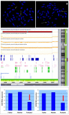
(a) Locus-specific FISH with N0002N02 (left) and N0418K11 (right) clones showing normal hybridization pattern (pink signals) on both chromosomes 7 (arrows). (b) SNP array analysis showing a deletion (red bar) of about 6.3 Mb at 7q21.13q21.3 proximal to DLX5 and DLX6 that encompasses 70 Refseq genes including DYNC1I1 (black arrow). (c) DLX5/6 expression calculated on RNA extracted from lymphoblastoid cell lines of the patient and his parents, showing a reduction in the patient’s sample (red arrows).
Figure 4
Figure 4
Detail of the distal portion of deletion (black line and arrow) and Hi-C map with TADs organization showing high grade of interactions (bright red dots) among the distal part of DYNC1I1 and DLX5/6 genes (dotted lines and circle). These data confirm that DYNC1I1 exons 15 and 17 act as enhancers of DLX5/6 by means of a physical interaction. The deletion includes the proximal TAD boundary and DYNC1I1, but not DLX5/6 genes. The Hi-C map was visualized on 3D Genome Browser, based on data provided from GM12868 cell line by Rao et al., 2014. (http://3dgenome.fsm.northwestern.edu/view.php; last access 6 June 2023).
Figure 5
Figure 5
Chromosomal alterations of previously reported cases [https://genome.ucsc.edu/ GRCh37/hg19 (accessed on 23 June 2023)] [6,20,21,27,28,29,30,31,32,33]. Deletions are represented in red, inversions in green and translocations in orange; rearrangements with a single breakpoint or breakpoints that are very close to each other are highlighted with an arrow. Note the position of DLX5/6 and that of eExons 15–17 of DYNC1I1. The SLC25A13 gene, included in some of the deletions, is correlated to an autosomal recessive disease.

Similar articles

Cited by

References

    1. Duijf P.H., van Bokhoven H., Brunner H.G. Pathogenesis of split-hand/split-foot malformation. Hum. Mol. Genet. 2003;12:51R–60R. doi: 10.1093/hmg/ddg090. - DOI - PubMed
    1. Sifakis S., Basel D., Ianakiev P., Kilpatrick M., Tsipouras P. Distal limb malformations: Underlying mechanisms and clinical associations. Clin. Genet. 2001;60:165–172. doi: 10.1034/j.1399-0004.2001.600301.x. - DOI - PubMed
    1. Ullah A., Gul A., Umair M., Irfanullah, Ahmad F., Aziz A., Wali A., Ahmad W. Homozygous sequence variants in the WNT10B gene underlie split hand/foot malformation. Genet. Mol. Biol. 2018;41:1–8. doi: 10.1590/1678-4685-gmb-2016-0162. - DOI - PMC - PubMed
    1. Sowińska-Seidler A., Socha M., Jamsheer A. Split-hand/foot malformation—Molecular cause and implications in genetic counseling. J. Appl. Genet. 2013;55:105–115. doi: 10.1007/s13353-013-0178-5. - DOI - PMC - PubMed
    1. Bernardini L., Palka C., Ceccarini C., Capalbo A., Bottillo I., Mingarelli R., Novelli A., Dallapiccola B. Complex rearrangement of chromosomes 7q21.13-q22.1 confirms the ectrodactyly-deafness locus and suggests new candidate genes. Am. J. Med. Genet. Part A. 2007;146A:238–244. doi: 10.1002/ajmg.a.32093. - DOI - PubMed

Supplementary concepts