

A Splicing Variant in RDH8 Is Associated with Autosomal Recessive Stargardt Macular Dystrophy
- PMID: 37628710
- PMCID: PMC10454646
- DOI: 10.3390/genes14081659
A Splicing Variant in RDH8 Is Associated with Autosomal Recessive Stargardt Macular Dystrophy
Abstract
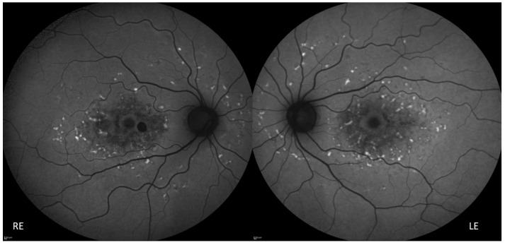
Stargardt macular dystrophy is a genetic disorder, but in many cases, the causative gene remains unrevealed. Through a combined approach (whole-exome sequencing and phenotype/family-driven filtering algorithm) and a multilevel validation (international database searching, prediction scores calculation, splicing analysis assay, segregation analyses), a biallelic mutation in the RDH8 gene was identified to be responsible for Stargardt macular dystrophy in a consanguineous Italian family. This paper is a report on the first family in which a biallelic deleterious mutation in RDH8 is detected. The disease phenotype is consistent with the expected phenotype hypothesized in previous studies on murine models. The application of the combined approach to genetic data and the multilevel validation allowed the identification of a splicing mutation in a gene that has never been reported before in human disorders.
Keywords: RDH8; Stargardt disease; all-trans-retinol dehydrogenase; macular dystrophy; retinal disease.
Conflict of interest statement
The authors declare no conflict of interest.
Figures


Similar articles
-
Whole exome sequencing identifies a novel splice-site mutation in IMPG2 gene causing Stargardt-like juvenile macular dystrophy in a north Indian family.Gene. 2022 Mar 30;816:146158. doi: 10.1016/j.gene.2021.146158. Epub 2022 Jan 4. Gene. 2022. PMID: 34990796
-
Targeted next-generation sequencing identifies ABCA4 mutations in Chinese families with childhood-onset and adult-onset Stargardt disease.Biosci Rep. 2021 Jun 25;41(6):BSR20203497. doi: 10.1042/BSR20203497. Biosci Rep. 2021. PMID: 33988224 Free PMC article.
-
Novel variants associated with Stargardt disease in Chinese patients.Gene. 2020 Sep 5;754:144890. doi: 10.1016/j.gene.2020.144890. Epub 2020 Jun 10. Gene. 2020. PMID: 32534057
-
Defective lipid transport and biosynthesis in recessive and dominant Stargardt macular degeneration.Prog Lipid Res. 2010 Oct;49(4):476-92. doi: 10.1016/j.plipres.2010.07.002. Epub 2010 Jul 13. Prog Lipid Res. 2010. PMID: 20633576 Free PMC article. Review.
-
Structure and function of ABCA4 and its role in the visual cycle and Stargardt macular degeneration.Prog Retin Eye Res. 2022 Jul;89:101036. doi: 10.1016/j.preteyeres.2021.101036. Epub 2021 Dec 23. Prog Retin Eye Res. 2022. PMID: 34954332 Review.
Cited by
-
Retinoid dynamics in vision: from visual cycle biology to retina disease treatments.Pharmacol Ther. 2025 Sep;273:108902. doi: 10.1016/j.pharmthera.2025.108902. Epub 2025 Jun 21. Pharmacol Ther. 2025. PMID: 40550364 Free PMC article. Review.
-
The Revolution of Genetic Diagnosis: An Example from Rare Disorders.Genes (Basel). 2024 Oct 15;15(10):1328. doi: 10.3390/genes15101328. Genes (Basel). 2024. PMID: 39457452 Free PMC article.
-
Evaluation of mesenchymal stem cells as an in vitro model for inherited retinal diseases.Front Cell Dev Biol. 2024 Nov 15;12:1455140. doi: 10.3389/fcell.2024.1455140. eCollection 2024. Front Cell Dev Biol. 2024. PMID: 39620144 Free PMC article. Review.
-
Scavenging of Cation Radicals of the Visual Cycle Retinoids by Lutein, Zeaxanthin, Taurine, and Melanin.Int J Mol Sci. 2023 Dec 29;25(1):506. doi: 10.3390/ijms25010506. Int J Mol Sci. 2023. PMID: 38203675 Free PMC article.
References
-
- Stargardt K. Über familiäre, progressive Degeneration in der Maculagegend des Auges. Albrecht Von Graefes Arch. Klin Ophthalmol. 1909;71:534–550. doi: 10.1007/BF01961301. - DOI
-
- Franceschetti A. A special form of tapetoretinal degeneration: Fundus flavimaculatus. Trans. Am. Acad. Ophthalmol. Otolaryngol. 1965;69:1048–1053. - PubMed
Publication types
MeSH terms
LinkOut - more resources
Full Text Sources
Molecular Biology Databases
