. 2023 Aug 8;24(16):12539.
doi: 10.3390/ijms241612539.

The Wound-Healing Activity of PEDOT-PSS in Animals

Affiliations

The Wound-Healing Activity of PEDOT-PSS in Animals

Yun-Lung Chung et al. Int J Mol Sci. .

Abstract

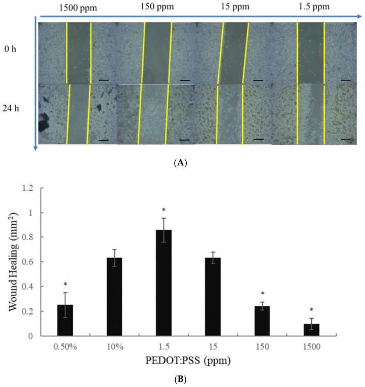
This study evaluated the wound-healing activity of a polymer, Poly(3,4-ethylenedioxythiophene):poly-(styrene sulfonate) (PEDOT: PSS), and determined its mechanism based on angiogenic activity in a full-thickness excision wound model in Spraque Dawley (SD) rats. Administering PEDOT: PSS (1.6) 1.5 ppm at a dose of 50 mg/kg/day significantly improved wound healing in the SD rats on the eleventh day after the incision was created. PEDOT: PSS-treated animals presented no anti-inflammatory skin effects; however, there was an increase in angiogenic behavior. VEGF was found to be significantly elevated in the PEDOT: PSS-treated groups seven days post-incision. However, only a higher concentration of PEDOT: PSS increased TGF-β1 expression within the same time frame. Our results showed that PEDOT: PSS enhances wound healing activity, mainly in terms of its angiogenic effects. In this paper, we describe the highly conductive macromolecular material PEDOT: PSS, which demonstrated accelerated wound-healing activity in the animal incision model. The results will further provide information regarding the application of PEDOT: PSS as a dressing for medical use.

Keywords: TGF-β1; VEGF; angiogenesis; wound-healing.


Conflict of interest statement

The authors declare no conflict of interests.

Figures

Figure 1
Figure 1
Cytotoxic effects of PEDOT: PSS on the human macrophage (RAW 264.7 (A) PEDOT-PSS (1.6) showed non-toxic effects in human macrophages (RAW 364.7). The cells were treated with different concentrations of PEDOT-PSS (1.6) (2.9, 5.9, 11.7, 23.4, 46.9, 93.8, 187.5, 375, 750, and 1500 ppm) for 24 h. These cells did not show apparent cell cytotoxicity. (B) PEDOT-PSS (2.5) and (C) PEDOT-PSS (5.0) at higher concentrations showed marked cell cytotoxicity. * p < 0.05, compared to the control group (treated with 10% FBS alone), according to a one-way analysis of variance (ANOVA).
Figure 2
Figure 2
Effect of PEDOT: PSS (1.6) on the wound healing process in SD rats. SD rats (n = 5) were administered with PEDOT: PSS (1.6) 1.5 ppm. It was administered to the control group with Vaseline ointment (100 mg per application/kg/day) and the PEDOT-PSS (1.6) experimental group at concentrations of 1.5 ppm for treatment (50 mg per application/day). Wound areas of the animals significantly reduced at day 11 and day 14 after the full-thickness excision compared to the control group (Day 0) (Table 1). Wound photographs were taken on days 3, 7, 11, and 14 after the incision. p < 0.05, compared to the control group.
Figure 3
Figure 3
Immunohistochemistry staining of PEDOT: PSS (1.6) on wound healing in the full-thickness-excision wound model in normal rats on day seven (VEGF stain). Skin wounds displayed slight to moderate/severe angiogenesis (arrow) in G1: Control (AC. Animal code: 1-2), G2 (DF. Animal code: 2-5), G3 (GI. Animal code: 3-2), and G4 (JL. Animal code: 4-2). The VEGF staining frequency scores were 2.3 ± 0.4; 3.5 ± 0.5; 2.6 ± 1.5; and 4.2 ± 0.7 in G1, G2, G3, and G4, respectively. Magnification: 40×, 100×, and 400×.
Figure 4
Figure 4
Immunohistochemistry staining of PEDOT: PSS (1.6) on wound healing in the full-thickness-excision wound model in normal rats on day 7 (TGF-β1 stain). Skin wounds showed an absence of TGF-β1 expression in the angiogenesis of blood vessels in G1: Control (AC. Animal code: 1-3); however, minimal TGF-β1 expression in the angiogenesis of blood vessels (arrow) was noted in G2 (DF. Animal code: 2-3) and G3 (GI. Animal code: 3-2), and slight expression was noted in G4 (JL. Animal code: 4-5). The TGF-β staining frequency scores were 0.0 ± 0.0; 0.3 ± 0.5; 0.2 ± 0.4; and 0.8 ± 0.7 in G1, G2, G3, and G4, respectively. Magnification: 40×, 100×, and 400×.
Figure 5
Figure 5
(A) PEDOT-PSS (1.6) 1.5 ppm markedly produced wound healing/closure in vitro. The experimental results showed significant wound healing after 24 h after wound healing in the macrophages (macrophage; RAW264.7) cells. The original magnification: 10×. And, the scale bar = 200 μM. (B) * p < 0.05 compared with the control group (according to one-way ANOVA).
Figure 6
Figure 6
Different particle sizes between PEDOT: PSS (1.6), PEDOT: PSS (2.5), and PEDOT: PSS (5.0).

References

    1. Keskin E.S., Keskin E.R., Ozturk M.B., Cakan D. The Effect of MMP-1 on Wound Healing and Scar For-mation. Aesthetic Plast. Surg. 2021;45:2973–2979. doi: 10.1007/s00266-021-02369-2. - DOI - PubMed
    1. Maver T., Maver U., Stana Kleinschek K., Smrke D.M., Kreft S. A review of herbal medicines in wound healing. Int. J. Dermatol. 2015;54:740–751. doi: 10.1111/ijd.12766. - DOI - PubMed
    1. Zhu S., Yu Y., Ren Y., Xu L., Wang H., Ling X., Jin L., Hu Y., Zhang H., Miao C., et al. The emerging roles of neutrophil extracellular traps in wound healing. Cell Death Dis. 2021;12:984. doi: 10.1038/s41419-021-04294-3. - DOI - PMC - PubMed
    1. Obagi Z., Damiani G., Grada A., Falanga V. Principles of Wound Dressings: A Review. Surg. Technol. Int. 2019;35:50–57. - PubMed
    1. Kloc M., Ghobrial R.M., Wosik J., Lewicka A., Lewicki S., Kubiak J.Z. Macrophage functions in wound healing. J. Tissue Eng. Regen. Med. 2019;13:99–109. doi: 10.1002/term.2772. - DOI - PubMed

LinkOut - more resources