. 2023 Aug 10;24(16):12626.
doi: 10.3390/ijms241612626.

A Novel Aniline Derivative from Peganum harmala L. Promoted Apoptosis via Activating PI3K/AKT/mTOR-Mediated Autophagy in Non-Small Cell Lung Cancer Cells

Affiliations

A Novel Aniline Derivative from Peganum harmala L. Promoted Apoptosis via Activating PI3K/AKT/mTOR-Mediated Autophagy in Non-Small Cell Lung Cancer Cells

Zhongnan Wu et al. Int J Mol Sci. .

Abstract

Non-small cell lung cancer (NSCLC) is a common clinical malignant tumor with limited therapeutic drugs. Leading by cytotoxicity against NSCLC cell lines (A549 and PC9), bioactivity-guided isolation of components from Peganum harmala seeds led to the isolation of pegaharoline A (PA). PA was elucidated as a structurally novel aniline derivative, originating from tryptamine with a pyrrole ring cleaved and the degradation of carbon. Biological studies showed that PA significantly inhibited NSCLC cell proliferation, suppressed DNA synthesis, arrested the cell cycle, suppressed colony formation and HUVEC angiogenesis, and blocked cell invasion and migration. Molecular docking and surface plasmon resonance (SPR) demonstrated PA could bind with CD133, correspondingly decreased CD133 expression to activate autophagy via inhibiting the PI3K/AKT/mTOR pathway, and increased ROS levels, Bax, and cleaved caspase-3 to promote apoptosis. PA could also decrease p-cyclinD1 and p-Erk1/2 and block the EMT pathway to inhibit NSCLC cell growth, invasion, and migration. According to these results, PA could inhibit NSCLC cell growth by blocking PI3K/AKT/mTOR and EMT pathways. This study provides evidence that PA has a promising future as a candidate for developing drugs for treating NSCLC.

Keywords: CD133; NSCLC; PI3K/AKT/mTOR; aniline derivative; apoptosis; autophagy.


Conflict of interest statement

The authors declare that they have no competing financial interests or personal relationships that may have any influence on the work reported in this paper.

Figures

Figure 1
Figure 1
PA (pegaharoline A) inhibited the proliferation of NSCLC cell lines (A549 and PC9) through promoting the production of ROS. (A) Chemical structure of PA. (B) The inhibition ratios of A549 and PC9 cells were detected by MTT assay at 48 h after treatment with various concentrations of PA. The DCFH-DA fluorescence probe was used for detecting ROS by using immunofluorescence and flow cytometry in PA-treated A549 cells (C) and PC9 cells (D) for 24 h, respectively. Scale bar 100 μm. Green dots in (C,D) (upper panel) indicated the ROS fluorescence-positive cells. Red parts in (C,D) (lower panel) indicated the rate of the ROS fluorescence-positive cells.
Figure 2
Figure 2
CD133 is overexpressed in lung cancer and strongly correlated with lung cancer “TNM” stage. (A) The amplification frequency of CD133 in lung cancer, and CD133 was strongly correlated with lung cancer “TNM” stage. Data were analyzed from cbioportal. (B) The expression of CD133 in pan-cancer. The arrows indicates lung adenocarcinoma. Data were analyzed from TCGA. (C) CD133 is overexpressed in lung cancer clinical tissue. Data were analyzed from “The Human Protein Atlas”. Scale bar, 100 μm. The arrows indicate overexpressed CD133.
Figure 3
Figure 3
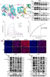
PA (pegaharoline A) inhibits CD133 expression to activate autophagy via blocking the PI3K/AKT/mTOR pathway. (A) Molecular docking diagram of ATP binding site between PA and CD133. The A549 and PC9 cells were treated with 3 μM PA at different times or treated for 24 h at different concentrations. The protein levels of CD133 were inhibited by PA both in A549 (B) and PC9 (C). (D) Analysis of the binding affinity of PA for CD133 protein by using SPR. SPR sensing map (D) of the interaction of CD133 with various concentrations of PA. The binding affinity curve (E) was obtained by fitting the single-site interaction model (different colored squares indicated the response unit (RU) of CD133 with the different concentrations (0.3125, 0.625, 1.25, 2.5, 5.0, 10.0 μM) of PA). (F) Immunofluorescence assay examined that treatment with PA for 24 h could promote the expression of autophagic protein LC3B both in A549 and PC9. Scale bar, 100 μm. (G,H) Western blotting assay detected the protein expression levels of P62, Atg5, Beclin 1, Lc3B, and PI3K/AKT/mTOR-pathway-related protein in A549 (G) and PC9 (H).
Figure 4
Figure 4
PA inhibits the proliferation of NSCLC cells by arresting cell cycle and promoting apoptosis. (A) The effects of PA on A549 cell cycle were detected by flow cytometry. The A549 cells were treated with PA for 24 h. (B) Statistical analysis of the A549 cell cycle. (C) The effects of PA on PC9 cell cycle were detected by using flow cytometry. The PC9 cells were treated with PA for 24 h. (D) Statistical analysis of the PC9 cell cycle. The results are representative of three independent experiments and are expressed as the mean ± SD. (E) The effect of PA on A549 cell apoptosis was detected by using flow cytometry. (F) Statistical analysis of A549 cell apoptosis. (G) The effect of PA on PC9 cell apoptosis was detected by using flow cytometry. (H) Statistical analysis of PC9 cell apoptosis. The results are representative of three independent experiments and are expressed as the mean ± SD. * p < 0.05, and ** p < 0.01 compared with the control group. The effects of PA on cell-cycle-related and apoptosis-related proteins in A549 cells (I) and PC9 cells (J) were detected by using Western blotting assay.
Figure 4
Figure 4
PA inhibits the proliferation of NSCLC cells by arresting cell cycle and promoting apoptosis. (A) The effects of PA on A549 cell cycle were detected by flow cytometry. The A549 cells were treated with PA for 24 h. (B) Statistical analysis of the A549 cell cycle. (C) The effects of PA on PC9 cell cycle were detected by using flow cytometry. The PC9 cells were treated with PA for 24 h. (D) Statistical analysis of the PC9 cell cycle. The results are representative of three independent experiments and are expressed as the mean ± SD. (E) The effect of PA on A549 cell apoptosis was detected by using flow cytometry. (F) Statistical analysis of A549 cell apoptosis. (G) The effect of PA on PC9 cell apoptosis was detected by using flow cytometry. (H) Statistical analysis of PC9 cell apoptosis. The results are representative of three independent experiments and are expressed as the mean ± SD. * p < 0.05, and ** p < 0.01 compared with the control group. The effects of PA on cell-cycle-related and apoptosis-related proteins in A549 cells (I) and PC9 cells (J) were detected by using Western blotting assay.
Figure 5
Figure 5
PA inhibited DNA synthesis, clone formation, and angiogenesis of HUVECs. (A) The effect of PA on DNA synthesis in A549 cells was detected by using EdU assay. The A549 cells were treated with PA for 24 h. Scale bar, 100 μm. (B) Statistical analysis of EdU-positive cells in A549 cells. (C) The effect of PA on DNA synthesis in PC9 cells was detected by using EdU assay. The PC9 cells were treated with PA for 24 h. Scale bar, 100 μm. (D) Statistical analysis of EdU-positive cells in PC9 cells. (E) The effect of PA on the colony formation of A549 cells was detected by colony formation assay. (F) Statistical analysis of the clone number of A549 cells after treatment with PA. (G) The effect of PA on the colony formation of PC9 cells. (H) Statistical analysis of the clone number of PC9 cells after treatment with PA. (I) The effect of PA on angiogenesis in HUVECs. Arrows: the angiogenesis-like structure. Scale bar, 100 μm. (J) The area ratio of vascular formation in HUVEC after treatment with PA. The results are representative of three independent experiments and are expressed as the mean ± SD. * p < 0.05, ** p < 0.01, and *** p < 0.001 compared with the control group.
Figure 6
Figure 6
PA inhibited the invasion and migration abilities of NSCLC cells by blocking EMT signal pathway. (A) The effects of PA on the invasion of A549 cells. Scale bar, 100 μm. (B) Statistical analysis of A549 cell invasive ability after treatment with PA. (C) The effects of PA on the invasion of PC9 cells. Scale bar, 100 μm. (D) Statistical analysis of PC9 cell invasive ability after treatment with PA. (E) The effects of PA on the migration of PC9 cells. Scale bar, 100 μm. (F) Statistical analysis of PC9 cell migration ability after treatment with PA. (G) The effects of PA on the migration of PC9 cells. Scale bar, 100 μm. (H) Statistical analysis of PC9 cell migration ability after treatment with PA. The effect of PA on the expression of EMT-related proteins in A549 (I) and PC9 (J) was detected by using Western blotting assay. Compared with the control group, the PA-treated group inhibited the EMT signal pathway by decreasing the expression of N-cadherin, Vimentin, Snail, while increasing the expression of E-cadherin. The results are representative of three independent experiments and are expressed as the mean ± SD. * p < 0.05 and ** p < 0.01 compared with the control group.
Figure 7
Figure 7
Mechanism schematic diagram of PA suppressing NSLC cells.

Similar articles

Cited by

References

    1. Chen W.Q., Li H., Sun K.X., Zheng R.S., He J. Report of cancer incidence and mortality in China, 2014. Chin. J. Oncol. 2018;40:5–13. doi: 10.21147/j.issn.1000-9604.2018.01.01. - DOI - PubMed
    1. Torre L.A., Siegel R.L., Jemal A. Lung Cancer and Personalized Medicine. Volume 893. Springer; Cham, Switzerland: 2016. Lung cancer statistics; pp. 1–19. (Part of Advances in Experimental Medicine and Biology Book Series (AEMB)). - DOI - PubMed
    1. Rossi A., La-Salvia A., Di-Maio M. Chemotherapy and intercalated gefitinib or erlotinib in the treatment of advanced non-small-cell lung cancer. Expert Rev. Respir. Med. 2017;11:171–180. doi: 10.1080/17476348.2017.1290526. - DOI - PubMed
    1. Ding P.N., Lord S.J., Gebski V., Links M., Bray V., Gralla R.J., Yang J.C., Lee C.K. Risk of treatment-related toxicities from EGFR tyrosine kinase inhibitors: A meta-analysis of clinical trials of gefitinib, erlotinib, and afatinib in advanced EGFR-mutated non-small cell lung cancer. J. Thorac. Oncol. 2017;12:633–643. doi: 10.1016/j.jtho.2016.11.2236. - DOI - PubMed
    1. Li J.N., Chen P.C., Wu Q.S., Gio L.B., Leong K.W., Chan K.L., Kwok H.F. A novel combination treatment of antiADAM17 antibody and erlotinib to overcome acquired drug resistance in non-small cell lung cancer through the FOXO3a/FOXM1 axis. Cell. Mol. Life Sci. 2022;79:614. doi: 10.1007/s00018-022-04647-x. - DOI - PMC - PubMed