Review
. 2023 Aug 10;24(16):12631.
doi: 10.3390/ijms241612631.

Smouldering Lesion in MS: Microglia, Lymphocytes and Pathobiochemical Mechanisms

Affiliations
Review

Smouldering Lesion in MS: Microglia, Lymphocytes and Pathobiochemical Mechanisms

Dániel Pukoli et al. Int J Mol Sci. .

Abstract

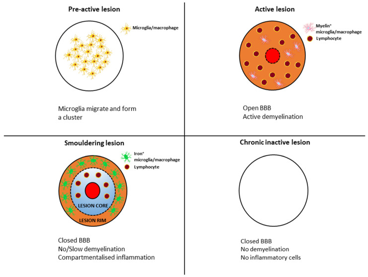
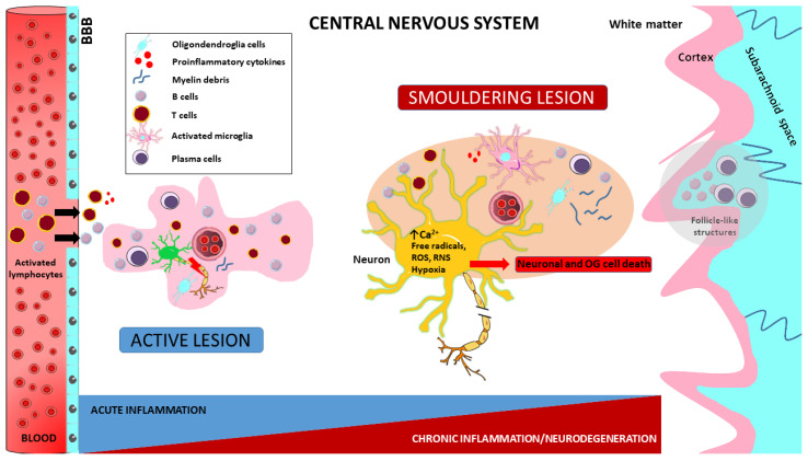
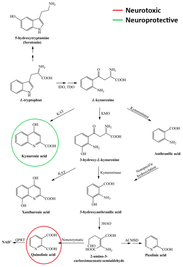
Multiple sclerosis (MS) is an immune-mediated, chronic inflammatory, demyelinating, and neurodegenerative disease of the central nervous system (CNS). Immune cell infiltration can lead to permanent activation of macrophages and microglia in the parenchyma, resulting in demyelination and neurodegeneration. Thus, neurodegeneration that begins with acute lymphocytic inflammation may progress to chronic inflammation. This chronic inflammation is thought to underlie the development of so-called smouldering lesions. These lesions evolve from acute inflammatory lesions and are associated with continuous low-grade demyelination and neurodegeneration over many years. Their presence is associated with poor disease prognosis and promotes the transition to progressive MS, which may later manifest clinically as progressive MS when neurodegeneration exceeds the upper limit of functional compensation. In smouldering lesions, in the presence of only moderate inflammatory activity, a toxic environment is clearly identifiable and contributes to the progressive degeneration of neurons, axons, and oligodendrocytes and, thus, to clinical disease progression. In addition to the cells of the immune system, the development of oxidative stress in MS lesions, mitochondrial damage, and hypoxia caused by the resulting energy deficit and iron accumulation are thought to play a role in this process. In addition to classical immune mediators, this chronic toxic environment contains high concentrations of oxidants and iron ions, as well as the excitatory neurotransmitter glutamate. In this review, we will discuss how these pathobiochemical markers and mechanisms, alone or in combination, lead to neuronal, axonal, and glial cell death and ultimately to the process of neuroinflammation and neurodegeneration, and then discuss the concepts and conclusions that emerge from these findings. Understanding the role of these pathobiochemical markers would be important to gain a better insight into the relationship between the clinical classification and the pathomechanism of MS.

Keywords: glutamate excitotoxicity; lymphocyte; microglia; mitochondrial dysfunction; multiple sclerosis; neurodegeneration; neuroinflammation; oxidative stress; slowly expanding lesion; smouldering lesion.


Conflict of interest statement

The authors have no conflict of interest to declare.

Figures

Figure 1
Figure 1
Transformation of typical lesion patterns in the course of multiple sclerosis.
Figure 2
Figure 2
Representation of smouldering lesions in MS. Iron—iron negative; SEL—slowly expanding lesion; PRL—paramagnetic rim lesion.
Figure 3
Figure 3
Pathogenetic implications in MS. Impaired autoreactive T-cell function is a consequence of Foxp3 T-cell dysregulation leading to T-cell maturation and proliferation induced by unidentified extrinsic or intrinsic antigens. The resulting activated T lymphocytes engage antigen-presenting B lymphocytes and induce B-cell maturation and differentiation into antibody-secreting plasma cells. Both B and T lymphocytes cross the blood–brain barrier (BBB) and enter the CNS. This results in damage to myelin, oligodendrocytes and prephagocytic neurons and the release of highly pro-inflammatory molecules. These, in turn, facilitate the migration of other monocytes and macrophages from the periphery to initiate phagocytosis. In the brain, meningeal B lymphocytes enhance inflammation through antigen presentation and antibody production, leading to cortical demyelination. Macrophages and microglia, which play a fundamental role in innate immunity, are also essential for disease progression by multiple mechanisms. Over the course of the disease, active lesions can evolve into smouldering lesions characterised by compartmentalised inflammation. These lesions consist of a thin rim of activated microglia interspersed with iron and myelin debris, with macrophages/microglia almost completely absent from their centres. In addition, meningeal follicles rich in B-cells are responsible for the subpial cortical demyelination and neuronal degeneration observed in both early and progressive forms of MS. Furthermore, progressive disease and increased neuronal transaction are associated with widespread and even overrepresented activated microglia or microglial nodules, which are already present in early non-lesional MS. Finally, the immune cells involved in these processes release a variety of neurotoxic substances, including antibodies, cytokines, free radicals and proteases. Activated macrophages and microglia release reactive oxygen species (ROS) and reactive nitrogen species (RNS), leading to mitochondrial damage, hypoxia with oligodendroglia (OG) and neuronal cell death.
Figure 4
Figure 4
Scheme of the kynurenine pathway. As an essential amino acid, tryptophan is mainly catabolised via the kynurenine pathway, leading to the production of neurotoxic and neuroprotective metabolites. 3-HAO—3-hydroxyanthranilate oxidase; ACMSD—α-amino-β-carboxymuconate-semialdehyde-decarboxylase; IDO—indoleamine 2,3-dioxygenase; KAT—kynurenine aminotransferase; KMO—kynurenine 3-monooxygenase; NAD+—nicotinamide adenine dinucleotide; QPRT—quinolinate phosphoribosyltransferase; TDO—tryptophan 2,3-dioxygenase.
Figure 5
Figure 5
Fe2+ (free iron) reacts through the Fenton reaction with hydrogen peroxide (H2O2), leading to the generation of highly reactive and damaging hydroxyl radicals (OH•). Highly reactive free hydroxyl radicals interact with molecules leading to the production of other free radicals, which then lead to oxidative stress-induced mitochondrial dysfunction, lipid peroxidation, DNA damage and, ultimately, cell dysfunction, and death.

Similar articles

Cited by

References

    1. Lassmann H., Bruck W., Lucchinetti C.F. The immunopathology of multiple sclerosis: An overview. Brain Pathol. 2007;17:210–218. doi: 10.1111/j.1750-3639.2007.00064.x. - DOI - PMC - PubMed
    1. Biström M., Jons D., Engdahl E., Gustafsson R., Huang J., Brenner N., Butt J., Alonso-Magdalena L., Gunnarsson M., Vrethem M., et al. Epstein–Barr virus infection after adolescence and human herpesvirus 6A as risk factors for multiple sclerosis. Eur. J. Neurol. 2020;28:579–586. doi: 10.1111/ene.14597. - DOI - PMC - PubMed
    1. Lublin F.D. New Multiple Sclerosis Phenotypic Classification. Eur. Neurol. 2014;72:1–5. doi: 10.1159/000367614. - DOI - PubMed
    1. Cree B.A., Hollenbach J.A., Bove R., Kirkish G., Sacco S., Caverzasi E., Bischof A., Gundel T., Zhu A.H., Papinutto N., et al. Silent progression in disease activity-free relapsing multiple sclerosis. Ann. Neurol. 2019;85:653–666. - PMC - PubMed
    1. Jonkman L.E., Soriano A.L., Amor S., Barkhof F., van der Valk P., Vrenken H., Geurts J.J.G. Can MS lesion stages be distinguished with MRI? A postmortem MRI and histopathology study. J. Neurol. 2015;262:1074–1080. doi: 10.1007/s00415-015-7689-4. - DOI - PMC - PubMed