. 2023 Aug 10;24(16):12637.
doi: 10.3390/ijms241612637.

Hybrids of Sterically Hindered Phenols and Diaryl Ureas: Synthesis, Switch from Antioxidant Activity to ROS Generation and Induction of Apoptosis

Affiliations

Hybrids of Sterically Hindered Phenols and Diaryl Ureas: Synthesis, Switch from Antioxidant Activity to ROS Generation and Induction of Apoptosis

Elmira Gibadullina et al. Int J Mol Sci. .

Abstract

The utility of sterically hindered phenols (SHPs) in drug design is based on their chameleonic ability to switch from an antioxidant that can protect healthy tissues to highly cytotoxic species that can target tumor cells. This work explores the biological activity of a family of 45 new hybrid molecules that combine SHPs equipped with an activating phosphonate moiety at the benzylic position with additional urea/thiourea fragments. The target compounds were synthesized by reaction of iso(thio)cyanates with C-arylphosphorylated phenols containing pendant 2,6-diaminopyridine and 1,3-diaminobenzene moieties. The SHP/urea hybrids display cytotoxic activity against a number of tumor lines. Mechanistic studies confirm the paradoxical nature of these substances which combine pronounced antioxidant properties in radical trapping assays with increased reactive oxygen species generation in tumor cells. Moreover, the most cytotoxic compounds inhibited the process of glycolysis in SH-SY5Y cells and caused pronounced dissipation of the mitochondrial membrane of isolated rat liver mitochondria. Molecular docking of the most active compounds identified the activator allosteric center of pyruvate kinase M2 as one of the possible targets. For the most promising compounds, 11b and 17b, this combination of properties results in the ability to induce apoptosis in HuTu 80 cells along the intrinsic mitochondrial pathway. Cyclic voltammetry studies reveal complex redox behavior which can be simplified by addition of a large excess of acid that can protect some of the oxidizable groups by protonations. Interestingly, the re-reduction behavior of the oxidized species shows considerable variations, indicating different degrees of reversibility. Such reversibility (or quasi-reversibility) suggests that the shift of the phenol-quinone equilibrium toward the original phenol at the lower pH may be associated with lower cytotoxicity.

Keywords: anticancer activity; apoptosis; cytotoxicity; electrochemical oxidation; glycolysis; mitochondrial membrane potential; molecular docking; quinone methides; sterically hindered phenol; urea.


Conflict of interest statement

The authors declare no conflict of interest.

Figures

Figure 1
Figure 1
(A) General approach to the creation of new types of anticancer agents based on sterically hindered phenols and the redox cycle of phenol-quinone methide. (B) The redox switch induced by oxidative stress in tumors. (C) Examples of diaryl ureas in drug design. (D) Acidification of the benzylic C-H group beside the phenolic OH is an important element of this structural design as it activates the O-H group. Note that if benzylic carbon is deprotonated, the O-H bond is significantly weakened. (E) Effects of substituents on C-H acidity at benzylic positions.
Scheme 1
Scheme 1
The synthesis of diarylmethylphosphonates containing sterically hindered phenols and (thio)urea moieties.
Figure 2
Figure 2
Molecular structures of compound 7a,b in crystals. Ellipsoids are shown with 50% probability.
Figure 3
Figure 3
1H-31P HMBC correlations of compound 5d.
Figure 4
Figure 4
Antioxidant potential of the tested compounds. (a) Inhibition of lipid peroxidation (LPO) in rat brain homogenates (2 mg/mL) initiated by Fe(II) ions (concentration of 500 µM) in the tested compounds at a concentration of 100 µM. (b) Concentration dependences of LPO inhibition for the studied compounds in the concentration range from 1 µM to 100 µM. (c) Oxygen Radical Absorbance Capacity (ORAC) of the studied compounds and ORAC values expressed as Trolox equivalent (TE). (d) DPPH antiradical activity of the compounds under study and DPPH values expressed as TE. The concentration of studied compounds and Trolox was 100 µM, the concentration of DPPH was 100 μM, and the concentration of AAPH was 12 µM. Statistical significance was assessed using one-way ANOVA and Dunnett’s multiple comparison tests, where **** p < 0.0001, *** p < 0.001 and ** p < 0.01 vs. control.
Figure 5
Figure 5
Quantitative analysis of the intracellular ROS level of compounds 11b and 17b at concentrations of 1/2 IC50 and IC50 in HuTu 80 cells using the CellROX® Deep Red flow cytometry kit. Bars show the average percent of fluorescent positive cells out of the total number of cells (n = 3). Statistical significance was assessed using one-way ANOVA and Dunnett’s multiple comparison tests, where **** p < 0.0001 and ** p < 0.01 vs. control.
Figure 6
Figure 6
Effect of synthesized compounds on the glycolytic profile of (ac) SH-SY5Y tumor cells and (d) calculated parameters of glycolytic function. The extracellular acidification rate (ECAR) is an indicator of glycolysis and shows the degree of extracellular acidification. The concentration of compounds was 100 µM, glucose—10 mM, oligomycin—1 µM и 2-fluoro-2-deoxy-D-glucose—25 mM. Data are presented as mean±error of the mean (n = 3). Statistical significance was assessed using one-way ANOVA and Dunnett’s multiple comparison tests, where **** p < 0.0001, *** p < 0.001, ** p < 0.01 and * p < 0.05 vs. control.
Figure 7
Figure 7
Docking of 11bR (a), 11bS (b) and 17bR (c) in allosteric activator pyruvate kinase PKM2. The indicated relationship with amino acids is shown for 11bR (polypeptide chain A: PHE 26—π–π stacking, polypeptide chain B: LYS 311—hydrogen bond, GLN 393—hydrogen bond), for 11bS (polypeptide chain A: TYR 390—hydrogen bond, polypeptide chain B: LYS 311—hydrogen bond, TYR 390—hydrogen bond, PHE 26—π–π stacking, LYS 311—π–cation interaction) and for 17bSR (polypeptide chain A: TYR 390—hydrogen bond, ARG 445—hydrogen bond, ARG 445—salt bridge, polypeptide chain B: LYS 311—hydrogen bond, PHE 26—π–π stacking).
Figure 8
Figure 8
Effect of the synthesized compounds on the membrane potential of rat liver mitochondria. (a) Kinetic lines of transmembrane potential changes at λex = 458 nm and λem = 590 nm. The concentration of the test substances was 100 µM. (b) Calculated parameters of % depolarization relative to control samples. Statistical significance was assessed using one-way ANOVA and Dunnett’s multiple comparison tests, where **** p < 0.0001 vs. control. (c) Concentration dependence of the effects of 11b and 17b on mitochondrial membrane potential. The concentration of the test substances was from 20 to 200 µM. Concentration of ions; Ca2+—25 µM. Mitochondria were energized by potassium succinate and rotenone. Data are presented as mean±error of the mean (n = 3).
Figure 9
Figure 9
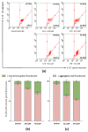
(a) Analysis of mitochondrial membrane potential using flow cytometry of HuTu 80 cells treated with compounds 11b and 17b at concentrations equal to ½ IC50 and IC50. (b,c) Quantitative determination of % cells with red aggregates and green monomers for 11b and 17b, respectively. Data are presented as ± SD (n = 3).
Figure 10
Figure 10
Cyclic voltammogram for the oxidation of SHPs. Conditions: 0.1 mM CH3CN (0.1 M Bu4NBF4); potentials vs. Ag/AgCl; work electrode: GC; scan rate: 0.1 V/s.
Figure 11
Figure 11
Differences in the transition back to the phenolic form after electrooxidation using data for compounds 13b (red) and 14b (black). Such differences may be helpful in identifying potential effective and non-effective compounds.
Figure 12
Figure 12
The SAR of a series of new diarylmethylphosphonates containing SHP and thiourea/urea moieties for cytotoxic activity in vitro against tumor lines.

References

    1. Liberti M.V., Locasale J.W. The Warburg Effect: How Does It Benefit Cancer Cells? Trends Biochem. Sci. 2016;41:211–218. doi: 10.1016/j.tibs.2015.12.001. - DOI - PMC - PubMed
    1. Panieri E., Santoro M.M. Ros Homeostasis and Metabolism: A Dangerous Liason in Cancer Cells. Cell Death Dis. 2016;7:e2253. doi: 10.1038/cddis.2016.105. - DOI - PMC - PubMed
    1. Czowski B.J., Romero-Moreno R., Trull K.J., White K.A. Cancer and PH Dynamics: Transcriptional Regulation, Proteostasis, and the Need for New Molecular Tools. Cancers. 2020;12:2760. - PMC - PubMed
    1. Yang W.Y., Breiner B., Kovalenko S.V., Ben C., Singh M., LeGrand S.N., Sang Q.X.A., Strouse G.F., Copland J.A., Alabugin I.V. C-Lysine Conjugates: PH-Controlled Light-Activated Reagents for Efficient Double-Stranded DNA Cleavage with Implications for Cancer Therapy. J. Am. Chem. Soc. 2009;131:11458–11470. doi: 10.1021/ja902140m. - DOI - PMC - PubMed
    1. Pérez-Herrero E., Fernández-Medarde A. The Reversed Intra- and Extracellular PH in Tumors as a Unified Strategy to Chemotherapeutic Delivery Using Targeted Nanocarriers. Acta Pharm. Sin. B. 2021;11:2243–2264. - PMC - PubMed