. 2023 Aug 15;24(16):12809.
doi: 10.3390/ijms241612809.

Beyond the Epidermal-Melanin-Unit: The Human Scalp Anagen Hair Bulb Is Home to Multiple Melanocyte Subpopulations of Variable Melanogenic Capacity

Affiliations

Beyond the Epidermal-Melanin-Unit: The Human Scalp Anagen Hair Bulb Is Home to Multiple Melanocyte Subpopulations of Variable Melanogenic Capacity

Cristina Casalou et al. Int J Mol Sci. .

Abstract

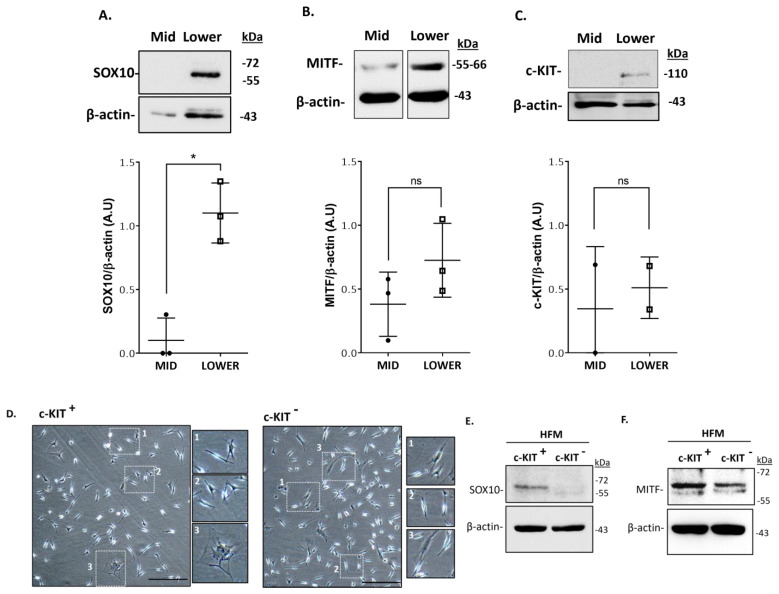
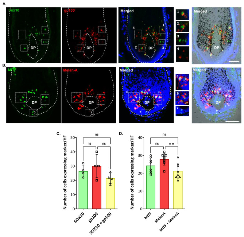
The visual appearance of humans is derived significantly from our skin and hair color. While melanin from epidermal melanocytes protects our skin from the damaging effects of ultraviolet radiation, the biological value of pigmentation in the hair follicle, particularly on the scalp, is less clear. In this study, we explore the heterogeneity of pigment cells in the human scalp anagen hair follicle bulb, a site conventionally viewed to be focused solely on pigment production for transfer to the hair shaft. Using c-KIT/CD117 microbeads, we isolated bulbar c-KIT-positive and c-KIT-negative melanocytes. While both subpopulations expressed MITF, only the c-KIT-positive fraction expressed SOX10. We further localized bulbar melanocyte subpopulations (expressing c-KIT, SOX10, MITF, and DCT) that exhibited distinct/variable expression of downstream differentiation-associated melanosome markers (e.g., gp100 and Melan-A). The localization of a second 'immature' SOX10 negative melanocyte population, which was c-KIT/MITF double-positive, was identified outside of the melanogenic zone in the most peripheral/proximal matrix. This study describes an approach to purifying human scalp anagen hair bulb melanocytes, allowing us to identify unexpected levels of melanocyte heterogeneity. The function of the more immature melanocytes in this part of the hair follicle remains to be elucidated. Could they be in-transit migratory cells ultimately destined to synthesize melanin, or could they contribute to the hair follicle in non-melanogenic ways?

Keywords: C-KIT; MITF; Melan-A; SOX10; bulbar melanocytes; follicular-melanin unit (FMU); hair follicle (HF); melanin.


Conflict of interest statement

The authors declare no conflict of interest.

Figures

Figure 1
Figure 1
Schematic representation of human anagen HF, indicating the distinct melanin units (EMU, FMUs) present in the upper, mid, and lower regions of HF, and the distinct subpopulations of melanocytes of each melanin unit. Magnified schematics of the lower bulb region of HF is shown.
Figure 2
Figure 2
Primary bulbar melanocytes of adult human scalp HF in primary culture (day 7), showing the presence of bipolar HF melanocytes (arrowheads), some intensely pigmented (yellow arrowheads), others fainter with low in melanin content (green arrowheads), together with poly-dendritic melanocytes with variable pigment content (blue arrows). Bar = 20 μm.
Figure 3
Figure 3
Isolation of melanocytes from adult human scalp HFs restricted to the mid-lower bulb region. Total protein extracts prepared from microdissected human follicles sectioned in mid and lower regions were analyzed by Western blot analysis for the expression of SOX10 (A), MITF (B), and c-KIT (C). Relative quantification of band intensities was made using β-actin or α-tubulin as loading controls. Data are the mean ± SD * p-value< 0.05; ns indicates not significant (non-parametric t-test; Wilcoxon). Further enrichment of melanocyte populations was made using CD117/c-KIT selection (MACS technology). (D) Phase contrast images representing the morphology of CD117-positive and CD117-negative cells at passage 1 (P1); attached bipolar and polydendritic cells (enlargements 1,2) and stromal cell (incl. fibroblast) contamination (enlargement 3). Scale bar 50 μm. SOX10 expression (E) was confirmed only in CD117-positive cell fraction, by Western blot analysis whereas MITF expression (F) was detected in both cell populations, (c-KIT+; c-KIT). β-actin was used as a loading control.
Figure 4
Figure 4
In situ localization of c-KIT expression in the proliferative bulb region of human adult scalp HF. c-KIT positive melanocytes were detected that lacked the differentiation marker, the (pre)-melanosomal protein gp100 (enlargements #1,2), while other c-KIT-positive melanocytes were already expressing gp100 (enlargements #3,4) (n = 3). Cell nuclei are indicated by DAPI staining. DP—dermal papilla. Scale bar 50 μm. The plot represents the quantification of the number of cells positive for each marker (gp100, green; c-KIT, red), and double-positive cells (n = 8; 4 donors). Data are the mean ± SD; ns indicates not significant (ordinary one-way ANOVA test).
Figure 5
Figure 5
c-KIT is variably co-expressed with MITF by melanocytes of the PP-FMU and M-FMU. Longitudinal cryosections of human adult scalp HFs were taken from the interior (A) and the exterior (B) regions and used to examine the expression of c-KIT and MITF (n = 2). (A) Magnifications 1–4 highlight the cellular co-localization of nuclear MITF with the cell membrane/cytoplasmatic c-KIT expression in the same cells of the melanogenically active region of HFs. (B) Melanocytes present at the peripheral proximal region (PP-FMU; enlargements #1–4) co-express c-KIT and MITF, whereas in the highly proliferative region of the hair bulb matrix (M-FMU) some melanocytes can be found that are only positive for MITF (enlargements #5–6). Cell nuclei are indicated by DAPI staining. Scale bar 50 μm. DP—dermal papilla. BF—Brightfield. (C,D) Quantification of the number of cells positive for each marker (c-KIT, red; MITF, green), and double-positive cells (n = 6; 3 donors in (A), n = 3, 3 donors in (B)). Data are the mean ± SD; ns indicates not significant (ordinary one-way ANOVA test).
Figure 6
Figure 6
Melanocyte expression of transcription factors MITF and SOX10 (in the proliferative bulb region of Human anagen HFs. The expression of SOX10 and MITF was assessed by IHC (A), together with melanocyte/melanosome markers gp100 and Melan-A (B). Enlargements #1–4 highlight the cellular localization of SOX10 and gp100, whereas #5–7 highlight the cellular localization of MITF and MelanA. Some melanocytes localized above the DP were SOX10 positive, but not yet making significant melanin (i.e., lacked gp100 expression; see enlargement #3), and others did not express SOX10 (enlargement #4). Cell nuclei are indicated by DAPI staining. DP—dermal papilla. Scale bar 50 μm. (C) Quantification of the number of cells positive for each marker (SOX10, green; gp100, red) and double-positive cells (n = 4; 3 donors). (D) Quantification of the number of cells positive for each marker (MITF, green; MelanA, red) and double-positive cells (n = 8; 3 donors). Data are the mean ± SD ** p-value < 0.01 and ns indicates not significant (ordinary one-way ANOVA test).
Figure 7
Figure 7
DCT was expressed by melanocytes in the melanogenically-active zone of the adult human scalp anagen bulb. (A) IHC analysis of DCT expression in the HF bulb region, where some melanocytes (red-only) expressed DCT, but expressed little or no gp100+ melanosomes (green). Enlargements #1–4 highlight DCT and gp100 cellular localization. DAPI -nuclear staining; DP—Dermal papilla. Scale bar 50 μm. (B) DCT was expressed in protein extracts of the melanogenic hair bulb, but not in protein extraction from the mid-region of HF, where O-FMU melanocytes reside (n = 3, 3 donors). IFNγ treatment (C) and UVB-induced stress (D) both upregulated DCT protein expression in the lower anagen hair bulb fractions, (n = 3, n = 2 respectively). β-actin and α-tubulin were used as loading controls. * p-value < 0.05 and ns indicates not significant (non-parametric t-test; Wilcoxon). (E) Quantification of the number of cells positive for each marker (DCT, green; gp100, red) and double-positive cells (n = 4; 3 donors). Data are the mean ± SD *** p-value < 0.001 * p-value < 0.05 and n.s. indicates not significant (ordinary one-way ANOVA test).

Similar articles

Cited by

References

    1. Paus R. Principles of Hair Cycle Control. J. Dermatol. 1998;25:793–802. doi: 10.1111/j.1346-8138.1998.tb02507.x. - DOI - PubMed
    1. Slominski A., Wortsman J., Płonka P.M., Schallreuter K.U., Paus R., Tobin D.J. Hair Follicle Pigmentation. J. Investig. Dermatol. 2005;124:13–21. doi: 10.1111/j.0022-202x.2004.23528.x. - DOI - PMC - PubMed
    1. Christoph T., Müller-Röver S., Audring H., Tobin D.J., Hermes B., Cotsarelis G., Rückert R., Paus R. The Human Hair Follicle Immune System: Cellular Composition and Immune Privilege. Br. J. Dermatol. 2000;142:862–873. doi: 10.1046/j.1365-2133.2000.03464.x. - DOI - PubMed
    1. Tobin D.J. The Cell Biology of Human Hair Follicle Pigmentation. Pigment. Cell Melanoma Res. 2010;24:75–88. doi: 10.1111/j.1755-148X.2010.00803.x. - DOI - PubMed
    1. Slominski A., Paus R. Melanogenesis Is Coupled to Murine Anagen: Toward New Concepts for the Role of Melanocytes and the Regulation of Melanogenesis in Hair Growth. J. Investig. Dermatol. 1993;101:90S–97S. - PubMed