Review
. 2023 Aug 16;24(16):12850.
doi: 10.3390/ijms241612850.

Extracellular Cysteine Proteases of Key Intestinal Protozoan Pathogens-Factors Linked to Virulence and Pathogenicity

Affiliations
Review

Extracellular Cysteine Proteases of Key Intestinal Protozoan Pathogens-Factors Linked to Virulence and Pathogenicity

Raúl Argüello-García et al. Int J Mol Sci. .

Abstract

Intestinal diseases caused by protistan parasites of the genera Giardia (giardiasis), Entamoeba (amoebiasis), Cryptosporidium (cryptosporidiosis) and Blastocystis (blastocystosis) represent a major burden in human and animal populations worldwide due to the severity of diarrhea and/or inflammation in susceptible hosts. These pathogens interact with epithelial cells, promoting increased paracellular permeability and enterocyte cell death (mainly apoptosis), which precede physiological and immunological disorders. Some cell-surface-anchored and molecules secreted from these parasites function as virulence markers, of which peptide hydrolases, particularly cysteine proteases (CPs), are abundant and have versatile lytic activities. Upon secretion, CPs can affect host tissues and immune responses beyond the site of parasite colonization, thereby increasing the pathogens' virulence. The four intestinal protists considered here are known to secrete predominantly clan A (C1- and C2-type) CPs, some of which have been characterized. CPs of Giardia duodenalis (e.g., Giardipain-1) and Entamoeba histolytica (EhCPs 1-6 and EhCP112) degrade mucin and villin, cause damage to intercellular junction proteins, induce apoptosis in epithelial cells and degrade immunoglobulins, cytokines and defensins. In Cryptosporidium, five Cryptopains are encoded in its genome, but only Cryptopains 4 and 5 are likely secreted. In Blastocystis sp., a legumain-activated CP, called Blastopain-1, and legumain itself have been detected in the extracellular medium, and the former has similar adverse effects on epithelial integrity and enterocyte survival. Due to their different functions, these enzymes could represent novel drug targets. Indeed, some promising results with CP inhibitors, such as vinyl sulfones (K11777 and WRR605), the garlic derivative, allicin, and purified amoebic CPs have been obtained in experimental models, suggesting that these enzymes might be useful drug targets.

Keywords: cysteine proteases; extracellular proteases; intestinal protozoa; papain-like proteases; pathogenicity; protists; virulence.


Conflict of interest statement

The authors declare no conflict of interest.

Figures

Figure 1
Figure 1
CPs from Giardia duodenalis trophozoites interact with multiple targets in the small intestine. Experimental models of trophozoites–epithelial cell interactions indicate that the parasite releases CPs, mainly cathepsin B-type, including Giardipain-1 (GL50803_14019), GL16160 and GL16779 and non-canonical CPs (e.g., VSP9B10A). This latter CP may serve, when expressed, to divert the immune system (1) or may cause damage to the epithelium due to a loss of cell–cell junction and cytotoxicity in the cell (2). Secreted CPs have been shown to degrade substrates, such as mucin 2, an important component of intestinal mucus (3), enabling trophozoite adhesion to microvilli. Direct damage to epithelial cell integrity by CPs may include disruption of the cytoskeletal microvillus-resident protein villin (4) and the disruption of tight junction proteins, such as ZO-1 and claudins, also involving adherens junction proteins including β-catenin or E-cadherin (5). Soluble elements of the innate immune response, including secretory IgA (6), produced by plasmatic B-cells, defensins (7) and IL-8 (8), both produced by epithelial cells, of which the latter works as neutrophil attractant, might be degraded by giardial CPs. These enzymes also provoke alterations in the microbiome of the small intestine, leading to dysbiosis (9), while bacterial translocation from the luminal to the intraepithelial compartment (10) may be promoted via the degradation of intercellular junctions by CPs as mentioned. Recent studies suggest that giardial CPs might be secreted after removal of N-terminal prodomain and inclusion into membrane-bound extracellular vesicles (EVs), mainly exosomes (green-filled circles).
Figure 2
Figure 2
Entamoeba histolytica CPs (EhCPs) are central to invasion, pathogenicity and immune evasion during intestinal and extraintestinal amoebiasis. Once the amoebic trophozoite reaches the mucus layer in the large intestine (a), EhCPs released in extracellular vesicles (EVs) can degrade the mucin backbone and secretory IgA antibodies (sIgA), which facilitates initiation of mucosal invasion (degradation is indicated by red rays). Intriguingly, the participation of EhCPs in the secretagogue capacity of the amoeba has also been described (activation is indicated by the blue rays). During trophozoite contact with the apical region of the intestinal epithelium (b), EhCPs degrade components of the extracellular matrix and villin in the apical region of enterocytes, eroding the epithelium and activating signaling pathways that lead to nuclear translocation of the transcription factor NF-κB, the inflammasome assembly and the expression of proinflammatory cytokines. The penetration of the amoeba through the epithelium (c) occurs in the intercellular spaces by EhCPs degradation of tight junctions, adhesion junctions and desmosomes components. At the same time, the parasite induces the death of enterocytes by trogocytosis and apoptosis, with EhCPs participating in the former. Since the amoeba is in the submucosa (d), the EhCPs can activate mast cells to produce IL-8, while they can degrade cytokines such as Pro-IL-18, or in contrast, activate them as in the case of Pro-IL-1B. At this point, EhCPs can also destroy intestinal nervous tissue (neurons/axons), affecting its physiology. During the invasion of the tissue, the amoeba also comes in contact with blood components (e), such as complement and IgG antibodies, which are degraded by EhCPs. At this point, in very sporadic cases and for reasons not yet understood, amoebae can migrate through the portal vein to the liver (f), where EhCPsbreak down hemoglobin to use iron. Finally, once in the liver (g), EhCPs contribute to tissue damage through the degradation of cell matrix components, enterocytes, and recruited immune cells, leading to the development of amoebic liver abscesses.
Figure 3
Figure 3
Protein structure models representing the Cryptopain family. The protein models of Cryptopain-1 (A), -2 (B), -3 (C), -4 (D) and -5 (E) were obtained using the I-Tasser server (https://zhanggroup.org/I-TASSER/; accessed dates: 28 February 2023, 3 March 2023, 22 and 28 July 2023), and the domains were identified with the InterPro platform (https://www.ebi.ac.uk/interpro/; accessed dates: 28 February 2023, 3 March 2023, 22 and 28 July 2023) and are indicated by colors at lower right. The catalytic triad Cys-His-Asn is displayed in ball-and-stick conformation and is magnified within dotted squares. Cryptopains 1-3 are cathepsin L-type and Cryptopains 4 and 5 are cathepsin B-type. From these analyses, Cryptopains 1 and 3 are predicted to be membrane-anchored (possess transmembrane domain), Cryptopain-2 is cytoplasmic (signal peptide and transmembrane domains absent) and Cryptopains 4 and 5 are secreted (possess signal peptide). Protein models were visualized and edited using the UCSF Chimera server v1.10.17.
Figure 4
Figure 4
Blastopain-1 and other cysteine proteases from Blastocystis. Possible role in pathogenesis and immune evasion. (A) Protein structure models of zymogen and active forms of a secreted, Cathepsin B (Blastopain-1) and legumain from Blastocystis sp. The catalytic triad Cys(red)-His(orange)-Asn (purple) is displayed in ball-and-stick conformation and is magnified within the dotted squares Protein domains were obtained from the InterPro platform and are colored as follows: signal peptides (SP) in magenta; papain-like signature (CatB) in green; cathepsin C prodomain (CatB) and auto-inhibitory C-terminal prodomain (legumain) in blue; hemoglobinase C13 signature (legumain) in cyan; and occluding loop (CatB) in brown. Models were obtained using the I-Tasser server from sequences with a.n. CBK25506-2 (CatB) and CBK21815-2 (legumain). (B) Proposed roles of cysteine proteases from Blastocystis. The cyst (C) form precedes the vegetative vacuolar (V) form that alternates with other entities (ameboid, granular) at intestinal lumen where cysteine proteases (CPs) may be secreted and causes effects at different levels: (1) parasites attached at intercellular junctions may release CPs that degrade junctional proteins such as ZO1- and claudins, promoting increased epithelial permeability; (2) CPs at intraepithelial compartment might induce upregulation of proinflammatory cytokines in Monocytes/Macrophages (Mϕ) and T lymphocytes; (3) epithelial cells exposed to CPs produce IL-8, a potent chemoattractant for Neutrophils and Polymorphonuclear cells; and (4) disruption of intercellular junctions along to a likely activation of Caspase-3 pathway may result in programmed cell death (apoptosis). Also, secreted CPs are able to degrade secretory IgA in vitro (5), an important effector in the mucosal system that has been observed at increased levels in symptomatic cases of Blastocystosis.
Figure 5
Figure 5
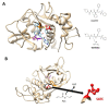
Mechanisms of inhibition of CPs from intestinal protozoa by synthetic and natural compounds. Protein models of secreted CPs were obtained by homology modeling using the Phyre2 server. (A) Blockade of the active site of EhCP1 by the vinyl sulfone K11777. Prediction model from SwissDock server (http://www.swissdock.ch/docking; accessed dates: 28 March 2023 and 11 July 2023) of a favored docking position (ΔG = −7.2436kCal/mol) of K11777 (in cyan, displayed in stick conformation) at the vicinity of the catalytic triad of EhCP1 displayed in ball-and-stick conformation (cysteine in red, histidine in orange and asparagine in purple). K11777 structure was obtained in SMILES format with further energy minimization using Avogadro suite v1.2. As reference, chemical structures of vinyl sulfone inhibitors K11777 and WRR483 are shown on the right. (B) Modification of active site cysteine from Giardipain-1 by allicin. The protein backbone shows the positions of catalytic residues that are displayed as described above. Upon interaction with allicin (ALC), the catalytic cysteine is converted into S-allylthiocysteine (SATC), which lacks nucleophilic nature as the cysteine thiol, thereby inactivating Giardipain-1. In this case, other thiol-disulfide exchange reactions could proceed with allosteric cysteines, perturbing enzyme activity. Protein models were visualized and edited using the UCSF Chimera server v1.10.17.

References

    1. Ximénez C., Cerritos R., Rojas L., Dolabella S., Morán P., Shibayama M., González E., Valadez A., Hernández E., Valenzuela O., et al. Human Amebiasis: Breaking the Paradigm? Int. J. Environ. Res. Public Health. 2010;7:1105–1120. doi: 10.3390/ijerph7031105. - DOI - PMC - PubMed
    1. Chalmers R.M. Chapter Sixteen—Cryptosporidium. In: Percival S.L., Yates M.V., Williams D.W., Chalmers R.M., Gray N.F., editors. Microbiology of Waterborne Diseases. 2nd ed. Academic Press; London, UK: 2014. pp. 287–326.
    1. Dogruman-Al F., Simsek Z., Boorom K., Ekici E., Sahin M., Tuncer C., Kustimur S., Altinbas A. Comparison of Methods for Detection of Blastocystis Infection in Routinely Submitted Stool Samples, and Also in IBS/IBD Patients in Ankara, Turkey. PLoS ONE. 2010;5:e15484. doi: 10.1371/journal.pone.0015484. - DOI - PMC - PubMed
    1. Argüello-García R., Carrero J.C., Ortega-Pierres G. Reference Module in Biomedical Sciences. Elsevier; Amsterdam, The Netherlands: 2020. Host Immune Responses Against Intestinal Unicellular Parasites and Their Role in Pathogenesis and Protection.
    1. Skotarczak B. Genetic Diversity and Pathogenicity of Blastocystis. Ann. Agric. Environ. Med. 2018;25:411–416. doi: 10.26444/aaem/81315. - DOI - PubMed