. 2023 Aug 18;24(16):12942.
doi: 10.3390/ijms241612942.

Heterodimerization of Chemoreceptors TAS1R3 and mGlu2 in Human Blood Leukocytes

Affiliations

Heterodimerization of Chemoreceptors TAS1R3 and mGlu2 in Human Blood Leukocytes

Lena Ball et al. Int J Mol Sci. .

Abstract

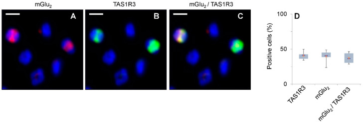
The expression of canonical chemosensory receptors of the tongue, such as the heteromeric sweet taste (TAS1R2/TAS1R3) and umami taste (TAS1R1/TAS1R3) receptors, has been demonstrated in many extra-oral cells and tissues. Gene expression studies have revealed transcripts for all TAS1 and metabotropic glutamate (mGlu) receptors in different types of immune cells, where they are involved, for example, in the chemotaxis of human neutrophils and the protection of T cells from activation-induced cell death. Like other class-C G protein-coupling receptors (GPCRs), TAS1Rs and mGlu receptors form heteromers within their families. Since mGlu receptors and TAS1R1/TAS1R3 share the same ligand, monosodium glutamate (MSG), we hypothesized their hitherto unknown heteromerization across receptor families in leukocytes. Here we show, by means of immunocytochemistry and co-IP/Western analysis, that across class-C GPCR families, mGlu2 and TAS1R3 co-localize and heterodimerize in blood leukocytes. Expressing the recombinant receptors in HEK-293 cells, we validated their heterodimerization by bioluminescence resonance energy transfer. We demonstrate MSG-induced, mGlu2/TAS1R3 heteromer-dependent gain-of-function and pertussis toxin-sensitive signaling in luminescence assays. Notably, we show that mGlu2/TAS1R3 is necessary and sufficient for MSG-induced facilitation of N-formyl-methionyl-leucyl-phenylalanine-stimulated IL-8 secretion in neutrophils, using receptor-specific antagonists. In summary, our results demonstrate mGlu2/TAS1R3 heterodimerization in leukocytes, suggesting cellular function-tailored chemoreceptor combinations to modulate cellular immune responses.

Keywords: BRET; ELISA; calcium fluorescence flow cytometry; chemosensory; fMLF; immunocytochemistry; taste receptors; transcript regulation.


Conflict of interest statement

The authors declare no conflict of interest.

Figures

Figure 1
Figure 1
GRM and TAS1R gene transcripts are expressed in human PMNs and T cells. (A,B) RT-qPCR demonstrates relative quantitative mRNA expression of GRM and TAS1R genes in human blood PMNs (A) and T cells (B). Data are shown as mean ± SD (n = 8, T cells; n = 9, PMNs). (C) Relative mRNA expression of GRMs and TAS1Rs in human PMNs after 24 h MSG stimulation. Data are shown as mean ± SEM (n = 8–15). Data were normalized to an averaged expression of two different reference genes (–ΔCT, GAPDH + ACTB). Transcript levels of GRM2 were significantly different compared to those of other GRMs and between TAS1R3 and the other TAS1Rs, as tested using a two-sided student’s t-test: (***) p ≤ 0.001; (**) p ≤ 0.01; (*) p ≤ 0.05. The dotted line indicates the fold change of 1, where there is no change in gene expression levels.
Figure 2
Figure 2
Two-color immunocytochemistry revealed co-expression of mGlu2 and TAS1R3 in a sub-population of isolated PMNs. (AC) Co-localization of mGlu2 and TAS1R3 in isolated PMNs (n = 6 blood samples, 3005 cells investigated). (A) anti-mGlu2 antibody and secondary antibody carrying fluorophore MFP631 (red); (B) anti-TAS1R3 antibody and secondary antibody carrying fluorophore MFP555 (green); (C) overlay of the signals in (A,B) (yellow). Cell nuclei were stained with Hoechst-33342 (blue). Original scale bars, 5 µm. (D) Box-whisker plots demonstrate the range of receptor positive cells. Lower and upper grey bars display 2nd and 3rd quartiles of data distribution; horizontal lines, median; X, data means.
Figure 3
Figure 3
Protein expression of mGlu2 and TAS1R3 in human blood PMNs. (A,B) Antibody validation of anti-mGlu2 (A) and anti-TAS1R3 antibody (B). Specificity of antibodies was tested by transfecting NxG cells with the respective recombinant receptors. Cell extracts were subjected to SDS-PAGE with an amount of 20 µg protein/lane. (C) Co-IP assay of mGlu2 and TAS1R3 in human blood PMNs. Cell lysates from PMNs (n = 3) were incubated with anti-mGlu2 (lane 1,) or anti-TAS1R3 antibody (lanes 2 + 3), attached to Dynabeads®. Lane 3, same as lane 2, but using 15,000× g (instead of 12,000× g) to remove membrane and unsolubilized receptor. For Western blot, whole cell lysates and immunoprecipitates were subjected to SDS-PAGE and were analyzed by immunoblotting using anti-TAS1R3 (lanes 1 + 5) or anti-mGlu2 antibodies (lanes 2–4). The predicted size of mGlu2 protein is ~110 kDa, for TAS1R3 protein ~ 97 kDa, indicated by the black arrows.
Figure 4
Figure 4
BRET detection of heterodimerization of recombinant mGlu2/TAS1R3 in HEK-293 cells. (A) Schematic overview of the NanoBRET™ system. Protein–protein interactions, in this case receptor dimerization, result in an energy transfer from a luminescent donor, the IL-6-NanoLuc® luciferase, to the IL-6-HaloTag® binding NanoBRET™ ligand, a fluorescent acceptor, whose excitation can be measured at 618 nm. (B) Results of the NanoBRET™-assay. The first mentioned receptor was always expressed out of vector pFN210A carrying the IL-6-HaloTag, the second one out of pNsecNLuc carrying the NanoLuc. Data are presented as the means ± SD of n = 4–11 independent experiments, normalized to positive control (PPI p53-pFN:MDM2-NL). Dashed line represents the highest value of the negative controls (black hatched bars). mBU, milli-BRET units. Significance of difference between receptor combinations and mock control was tested using a two-sided student’s t-test: (***) p ≤ 0.001; (**) p ≤ 0.01.
Figure 5
Figure 5
mGlu2 and TAS1R3 show gain-of-function in transfected HEK-293 cells. (A) Schematic overview of real-time cAMP luminescence-based test cell system. An agonist–receptor interaction results in an activation of the Gαi-subunit, which typically inhibits an adenylyl cyclase. A stimulation by forskolin, an adenylyl cyclase activator, leads to an increase in intracellular cAMP, synthesized by an adenylyl cyclase. A simultaneous stimulation of agonist and forskolin leads to an agonist concentration-dependent attenuation of forskolin-induced increase in intracellular cAMP. cAMP binds to a genetically modified luciferase and the emission of light was detected by the GloMax® Discover system (Promega, Madison, WI, USA). (B) Experimental procedure. After simultaneous stimulation of the cells with an agonist and forskolin, RLU values were measured for 21 min, until a plateau was detected, where lower RLU values were obtained for the agonist in comparison to the solvent control. Each data set was normalized to its maximum luminescence value leading to a concentration response curve where IC50 values were calculated. (C) Concentration response curves for MSG in transfected HEK-293 cells. Data are the mean ± SD (n = 3, in triplicates). RLU, relative luminescence units. Significance of difference between mGlu2 and mGlu2/TAS1R3 was tested using the two-sided student’s t-test: (*) p ≤ 0.05.
Figure 6
Figure 6
MSG via heteromeric mGlu2/TAS1R3 facilitated an fMLF-induced IL-8 secretion in isolated PMNs in vitro. (A) 4 h fMLF-induced IL-8 secretion in isolated PMNs, after 2 h pre-stimulation with 50 µM MSG or RPMI-treatment, in the absence or presence of mGlu2-specific antagonist 1 and/or TAS1R3-specific antagonist Lactisole. Changes of IL-8 concentration are normalized to RPMI-treated samples without MSG (“solvent”, n = 6–13). Data are shown as mean ± SEM. Significance of difference between solvent and MSG pre-stimulated cells was tested using a two-sided student’s t-test: (***) p ≤ 0.001; (**) p ≤ 0.01; (*) p ≤ 0.05. (B) Maximum fMLF-induced IL-8 concentrations in pg/mL in presence and absence of MSG pre-stimulus or the respective receptor-specific antagonists. Data are the mean ± SEM (n = 6–13). Significance of difference was tested using the one-sided student’s t-test: (**) p ≤ 0.01; (*) p ≤ 0.05.
Figure 7
Figure 7
MSG and the mGlu2-receptor agonist LY379268 increased intracellular Ca2+ in isolated PMNs. Shown are the number of Fluo-4-positive cells after a 2 h incubation with MSG or the mGlu2 receptor agonist LY379268 in the presence or absence of mGluR2 antagonist 1. Solvent control (0.1% DMSO) was subtracted from each measurement. Data are expressed as mean ± SEM (n = 4). The significance of differences was tested using the one-sided student’s t-test: (**) p ≤ 0.01; (*) p ≤ 0.05, n.s., not significant.

Similar articles

References

    1. Milligan G., Ward R.J., Marsango S. GPCR homo-oligomerization. Curr. Opin. Cell Biol. 2019;57:40–47. doi: 10.1016/j.ceb.2018.10.007. - DOI - PMC - PubMed
    1. Ferre S., Casado V., Devi L.A., Filizola M., Jockers R., Lohse M.J., Milligan G., Pin J.P., Guitart X. G protein-coupled receptor oligomerization revisited: Functional and pharmacological perspectives. Pharmacol. Rev. 2014;66:413–434. doi: 10.1124/pr.113.008052. - DOI - PMC - PubMed
    1. Lohse M.J. Dimerization in GPCR mobility and signaling. Curr. Opin. Pharmacol. 2010;10:53–58. doi: 10.1016/j.coph.2009.10.007. - DOI - PubMed
    1. Gurevich V.V., Gurevich E.V. GPCR monomers and oligomers: It takes all kinds. Trends Neurosci. 2008;31:74–81. doi: 10.1016/j.tins.2007.11.007. - DOI - PMC - PubMed
    1. Borroto-Escuela D.O., Brito I., Romero-Fernandez W., Di Palma M., Oflijan J., Skieterska K., Duchou J., Van Craenenbroeck K., Suarez-Boomgaard D., Rivera A., et al. The G protein-coupled receptor heterodimer network (GPCR-HetNet) and its hub components. Int. J. Mol. Sci. 2014;15:8570–8590. doi: 10.3390/ijms15058570. - DOI - PMC - PubMed

MeSH terms

LinkOut - more resources