. 2023 Aug 21;24(16):13012.
doi: 10.3390/ijms241613012.

Unveiling the Link: Exploring Mitochondrial Dysfunction as a Probable Mechanism of Hepatic Damage in Post-Traumatic Stress Syndrome

Affiliations

Unveiling the Link: Exploring Mitochondrial Dysfunction as a Probable Mechanism of Hepatic Damage in Post-Traumatic Stress Syndrome

Marina V Kondashevskaya et al. Int J Mol Sci. .

Abstract

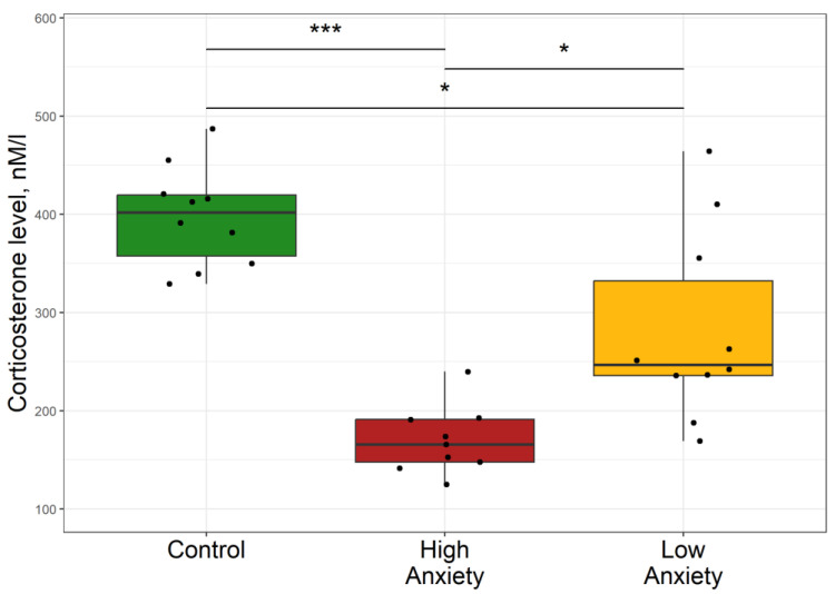
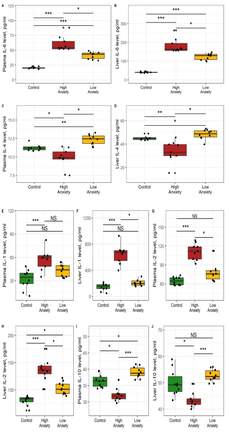
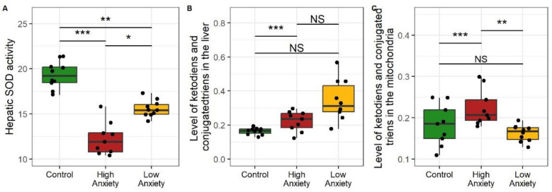
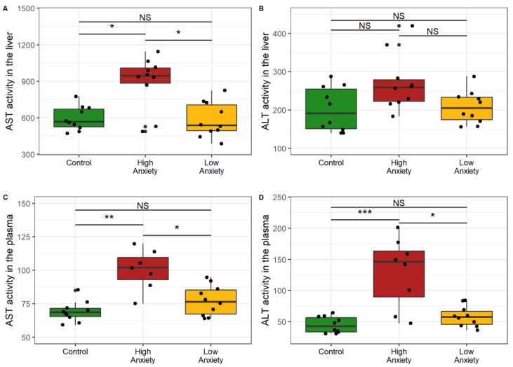
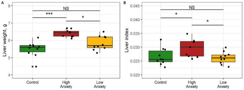
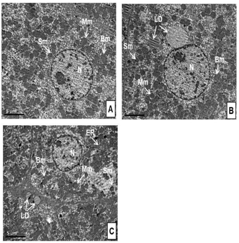
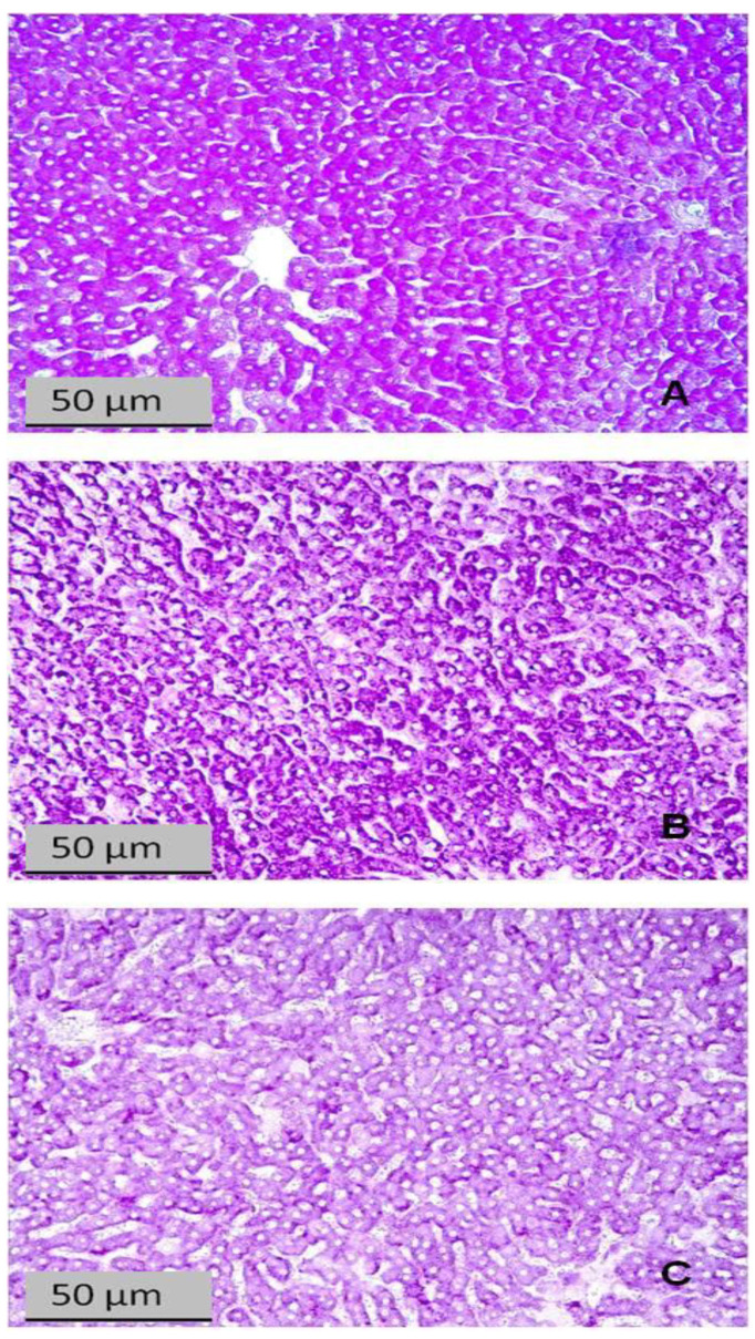
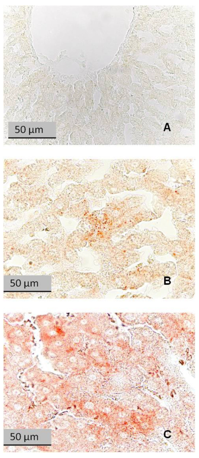
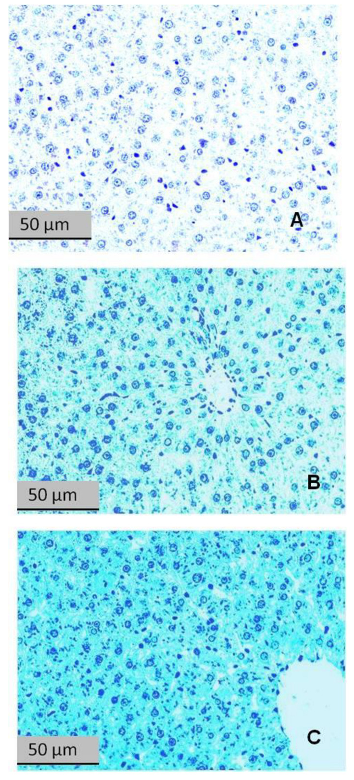
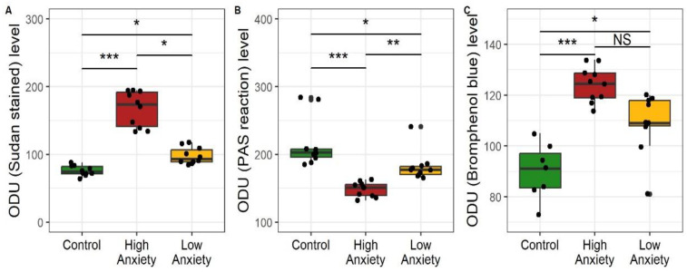
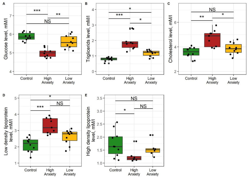
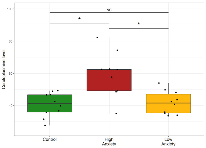
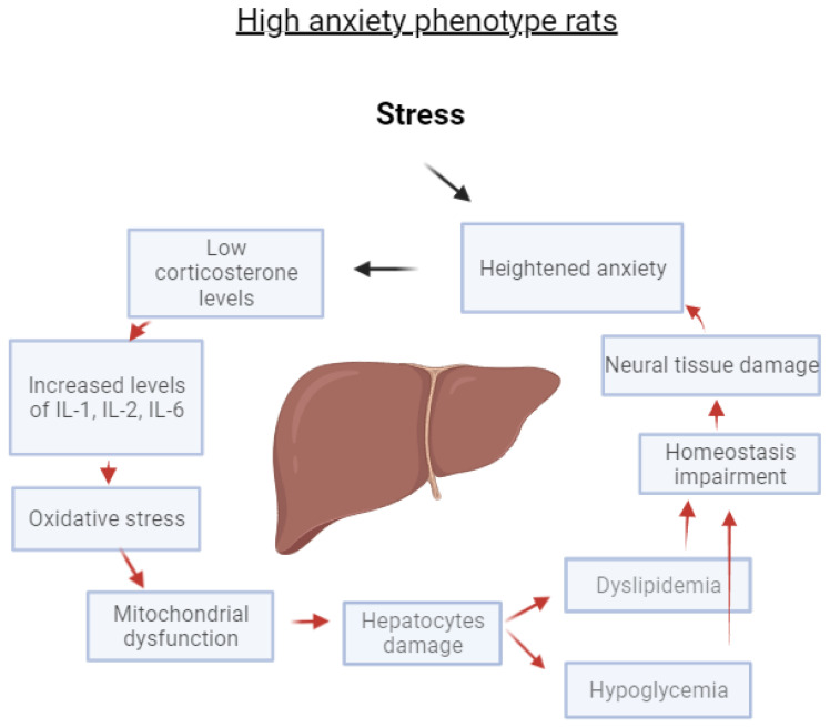
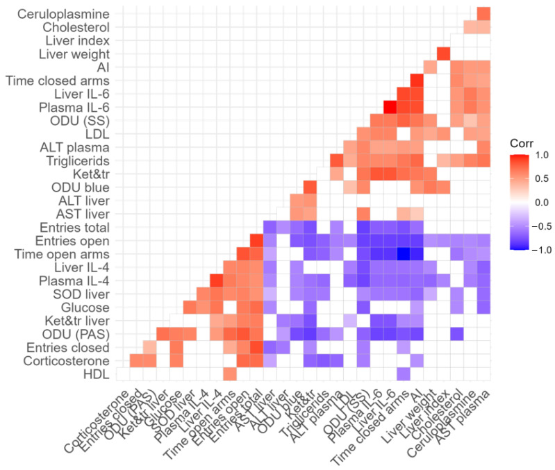
PTSD is associated with disturbed hepatic morphology and metabolism. Neuronal mitochondrial dysfunction is considered a subcellular determinant of PTSD, but a link between hepatic mitochondrial dysfunction and hepatic damage in PTSD has not been demonstrated. Thus, the effects of experimental PTSD on the livers of high anxiety (HA) and low anxiety (LA) rats were compared, and mitochondrial determinants underlying the difference in their hepatic damage were investigated. Rats were exposed to predator stress for 10 days. Then, 14 days post-stress, the rats were evaluated with an elevated plus maze and assigned to HA and LA groups according to their anxiety index. Experimental PTSD caused dystrophic changes in hepatocytes of HA rats and hepatocellular damage evident by increased plasma ALT and AST activities. Mitochondrial dysfunction was evident as a predominance of small-size mitochondria in HA rats, which was positively correlated with anxiety index, activities of plasma transaminases, hepatic lipids, and negatively correlated with hepatic glycogen. In contrast, LA rats had a predominance of medium-sized mitochondria. Thus, we show links between mitochondrial dysfunction, hepatic damage, and heightened anxiety in PTSD rats. These results will provide a foundation for future research on the role of hepatic dysfunction in PTSD pathogenesis.

Keywords: anxiety; cytokines; hepatocytes; inflammation; liver; mitochondria; oxidative stress; phenotypes; post-traumatic stress disorder; rats.


Conflict of interest statement

The authors declare no conflict of interest. The funding agencies had no role in the design of the study; in the collection, analyses, or interpretation of data; in the writing of the manuscript, or in the decision to publish the manuscript.

Figures

Figure 1
Figure 1
Differences in plasma corticosterone concentrations between the control, low anxiety, and high anxiety groups of rats. In this and in similar figures, the boxes include the middle 50% of the data, i.e., from the 25th to the 75th percentile, with the median value shown by the horizontal line. The whiskers include data that fall within 1.5 times the interquartile range. p-values were determined by non-parametric analysis. Control n = 10; High Anxiety n = 9; Low Anxiety n = 10. * p < 0.05, *** p < 0.001.
Figure 2
Figure 2
Effects of PS on cytokine concentrations in plasma and liver. * p < 0.05, ** p < 0.01, *** p < 0.001, NS, not significant. (A) plasma IL-6; (B) liver IL-6; (C) plasma IL-4; (D) liver IL-4; (E) plasma IL-1; (F) liver, IL-1; (G) plasma IL-2; (H) liver IL-2; (I) plasma IL-10; (J) liver IL-10.
Figure 3
Figure 3
Oxidative stress in the liver as reflected by hepatic superoxide dismutase (SOD), ketodienes, and conjugated trienes. * p < 0.05, ** p < 0.01, *** p < 0.001, NS, not significant. (A) Hepatic SOD activity; (B) Hepatic concentrations of ketodienes and conjugated trienes; (C) Mitochondrial concntrations of ketodienes and conjugated trienes. SOD activity is expressed in units min−1 mg protein; ketodienes and conjugated trienes are expressed as oxidation indices E278/220.
Figure 4
Figure 4
AST and ALT activity in plasma and liver. * p < 0.05, ** p < 0.01, *** p < 0.001, NS, not significant. (A) AST activity in liver; (B) ALT activity in liver; (C) AST activity in plasma; (D) ALT activity in plasma.
Figure 5
Figure 5
Liver weight and weight index values in control and stress-exposed rats. * p < 0.05, *** p < 0.001, NS, not significant. (A) Liver weight; (B) Liver index.
Figure 6
Figure 6
Ultrastructure of liver cells of control rats (A), LA rats (B), and HA (C) rats. N, nucleus; Sm, small mitochondria; Mm, medium mitochondria; Bm, big mitochondria; ER, endoplasmic reticulum; LD, lipid droplets. Bar = 5 µm.
Figure 7
Figure 7
Liver sections stained with Schiff reagent for glycogen (PAS reaction). (A) Control rat; (B) LA rat; (C) HA rat.
Figure 8
Figure 8
Liver sections stained with Sudan III for lipids. (A) Control rat; (B) LA rat; (C) HA rat. Reddish-orange spots in hepatocytes are neutral lipids.
Figure 9
Figure 9
Liver sections stained with bromophenol blue for protein content. (A) Control rat; (B) LA rat; (C) HA rat.
Figure 10
Figure 10
Optical density (optical density units, ODU) of liver sections stained for (A) neutral fats, Sudan III, (B) glycogen, periodic acid-Schiff (PAS) reaction, (C) protein, bromophenol blue. * p < 0.05, ** p < 0.01, *** p < 0.001, NS, not significant.
Figure 11
Figure 11
Glucose (A), triglycerides (B), cholesterol (C), and low-density (D) and high-density (E) lipoprotein concentrations in blood of control, HA, and LA rats. * p < 0.05, ** p < 0.01, *** p < 0.001, NS, not significant.
Figure 12
Figure 12
Ceruloplasmin in control and stress-exposed rats. * p < 0.05, NS, not significant.
Figure 13
Figure 13
A positive feedback loop explains the interrelations between liver damage and anxiety in HA rats exposed to experimental PS.
Figure 14
Figure 14
Correlation matrix showing the relationship between measured variables in PS-exposed rats. Respective r values are indicated by the color bar below the matrix. p < 0.05 for all colored correlations. Details are shown in the Supplementary File.

Similar articles

Cited by

References

    1. Tanaka M., Szabó Á., Vécsei L. Preclinical modeling in depression and anxiety: Current challenges and future research directions. Adv. Clin. Exp. Med. 2023;32:505–509. doi: 10.17219/acem/165944. - DOI - PMC - PubMed
    1. Kerbage H., Bazzi O., El Hage W., Corruble E., Purper-Ouakil D. Early interventions to prevent post-traumatic stress disorder in youth after exposure to a potentially traumatic event: A scoping review. Healthcare. 2022;28:818. doi: 10.3390/healthcare10050818. - DOI - PMC - PubMed
    1. Smagin D., Kovalenko I., Galyamina A., Belozertseva I., Tamkovich N., Baranov K.O., Kudryavtseva N. Chronic lithium treatment affects anxious behaviors and the expression of serotonergic genes in midbrain raphe nuclei of defeated male mice. Biomedicines. 2021;22:1293. doi: 10.3390/biomedicines9101293. - DOI - PMC - PubMed
    1. Fava G., McEwen B., Guidi J., Gostoli S., Offidani E., Sonino N. Clinical characterization of allostatic overload. Psychoneuroendocrinology. 2019;108:94–101. doi: 10.1016/j.psyneuen.2019.05.028. - DOI - PubMed
    1. Stankiewicz A., Swiergiel A., Lisowski P. Epigenetics of stress adaptations in the brain. Brain Res. Bull. 2013;98:76–92. doi: 10.1016/j.brainresbull.2013.07.003. - DOI - PubMed