

Position of the ISTENT Inject® Trabecular Micro-Bypass System Visualized with the NIDEK GS-1 Gonioscope-A Postoperative Analysis
- PMID: 37629213
- PMCID: PMC10455890
- DOI: 10.3390/jcm12165171
Position of the ISTENT Inject® Trabecular Micro-Bypass System Visualized with the NIDEK GS-1 Gonioscope-A Postoperative Analysis
Abstract
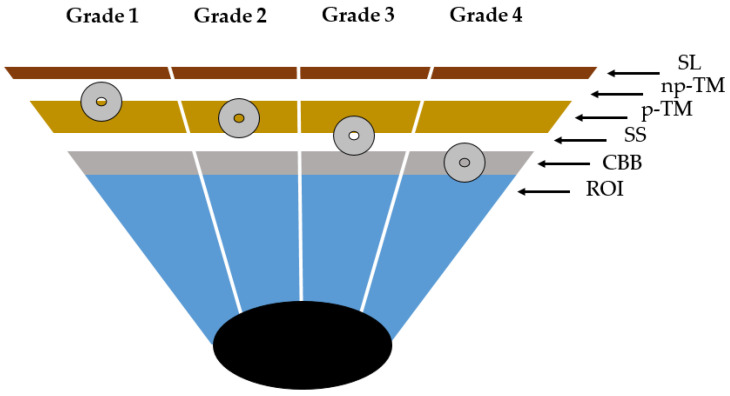
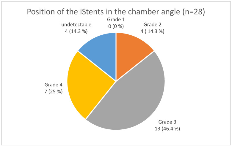
Glaucoma is one of the leading causes of irreversible blindness globally and is characterized by the gradual loss of retinal ganglion cells. The primary risk factor for the development and progression of glaucoma is increased intraocular pressure (IOP). Numerous surgical interventions exist to lower IOP should conservative therapy fail. One trend in recent years has been minimally invasive glaucoma surgery (MIGS) as an alternative to traditional methods. The ISTENT inject® is an ab interno trabecular micro-bypass implant designed to be implanted through the trabecular meshwork into the Schlemm's canal to lower IOP. The aim of the study was the postoperative visualization and description of the positioning of the ISTENT inject® using automated circumferential goniophotography. Patients with symptomatic cataracts and mild to moderate primary open-angle glaucoma (POAG), pseudoexfoliation glaucoma (PEX), and pigment-dispersion glaucoma were included who underwent combined cataract surgery with the ISTENT inject® and received postoperative automated gonioscopy with the NIDEK Gonioscope GS-1 to visualize the location of the implant. Twenty-four implants of 14 eyes in 11 patients could be visualized. Out of the implants, 14.3% were in the trabecular meshwork, 46.4% were at the border between the trabecular meshwork and scleral spur, 25% were below the trabecular meshwork, and 14.3% of the implants were not detectable in the gonioscopy. In the overall cohort, a statistically significant IOP reduction was found over the 12-month postoperative observation period. Even in three eyes, in each of which both stents were located below the trabecular meshwork, an IOP reduction over 12 months was observed compared to the baseline IOP. In this study, vertical two-dimensional positioning of the ISTENT inject® was performed for the first time using NIDKE GS-1 automated 360° goniophotography. The method is suitable for postoperative visualization, control, and documentation of positioning after ISTENT inject® implantation. Further studies are needed to analyze the correlation between positioning of the ISTENT inject® in the chamber angle and postoperative IOP reduction.
Keywords: MIGS; glaucoma; gonioscopy.
Conflict of interest statement
The authors declare no conflict of interest.
Figures
Similar articles
-
Ab interno trabecular bypass surgery with iStent for open-angle glaucoma.Cochrane Database Syst Rev. 2019 Mar 28;3(3):CD012743. doi: 10.1002/14651858.CD012743.pub2. Cochrane Database Syst Rev. 2019. PMID: 30919929 Free PMC article.
-
A Prospective Analysis of iStent Inject Microstent Positioning: Schlemm Canal Dilatation and Intraocular Pressure Correlations.J Glaucoma. 2019 Jul;28(7):613-621. doi: 10.1097/IJG.0000000000001273. J Glaucoma. 2019. PMID: 31058666 Clinical Trial.
-
One-Year Comparative Evaluation of iStent or iStent inject Implantation Combined with Cataract Surgery in a Single Center.Adv Ther. 2019 Oct;36(10):2797-2810. doi: 10.1007/s12325-019-01067-5. Epub 2019 Aug 22. Adv Ther. 2019. PMID: 31440981 Free PMC article.
-
Prospective Evaluation of Two iStent® Trabecular Stents, One iStent Supra® Suprachoroidal Stent, and Postoperative Prostaglandin in Refractory Glaucoma: 4-year Outcomes.Adv Ther. 2018 Mar;35(3):395-407. doi: 10.1007/s12325-018-0666-4. Epub 2018 Feb 23. Adv Ther. 2018. PMID: 29476443 Free PMC article.
-
iStent trabecular micro-bypass stent for open-angle glaucoma.Clin Ophthalmol. 2014 Sep 23;8:1937-45. doi: 10.2147/OPTH.S45920. eCollection 2014. Clin Ophthalmol. 2014. PMID: 25284980 Free PMC article. Review.
Cited by
-
iStent insertion orientation and impact on trabecular meshwork motion resolved by optical coherence tomography imaging.J Biomed Opt. 2024 Jul;29(7):076008. doi: 10.1117/1.JBO.29.7.076008. Epub 2024 Jul 27. J Biomed Opt. 2024. PMID: 39070082 Free PMC article.
-
Minimally Invasive Glaucoma Surgery Procedure in the Human Eye. A Fluid Structure Interaction Study.Int J Numer Method Biomed Eng. 2025 Jul;41(7):e70062. doi: 10.1002/cnm.70062. Int J Numer Method Biomed Eng. 2025. PMID: 40637285 Free PMC article.
-
Visualization of the Postoperative Position of the Hydrus® Microstent Using Automatic 360° Gonioscopy.J Clin Med. 2024 Sep 9;13(17):5333. doi: 10.3390/jcm13175333. J Clin Med. 2024. PMID: 39274546 Free PMC article.
References
-
- Gazzard G., Konstantakopoulou E., Garway-Heath D., Adeleke M., Vickerstaff V., Ambler G., Hunter R., Bunce C., Nathwani N., Barton K. Laser in Glaucoma and Ocular Hypertension (LiGHT) Trial: Six-Year Results of Primary Selective Laser Trabeculoplasty versus Eye Drops for the Treatment of Glaucoma and Ocular Hypertension. Ophthalmology. 2023;130:139–151. doi: 10.1016/j.ophtha.2022.09.009. - DOI - PubMed
-
- U.S. Food & Drug Administration Premarket Approval (PMA) [(accessed on 1 July 2023)]; Available online: https://www.accessdata.fda.gov/scripts/cdrh/cfdocs/cfpma/pma.cfm?id=P170043.
LinkOut - more resources
Full Text Sources
