

Evaluation of Retinal Blood Flow in Patients with Monoclonal Gammopathy Using OCT Angiography
- PMID: 37629268
- PMCID: PMC10456010
- DOI: 10.3390/jcm12165227
Evaluation of Retinal Blood Flow in Patients with Monoclonal Gammopathy Using OCT Angiography
Abstract
Background: Monoclonal gammopathy (MG) is characterized by monoclonal protein overproduction, potentially leading to the development of hyperviscosity syndrome.
Objective: To assess retinal circulation using optical coherence tomography angiography (OCTA) parameters in patients with monoclonal gammopathy.
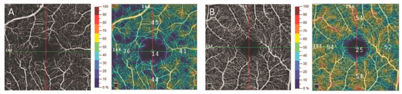
Methods: OCTA measurements were performed using the Optovue AngioVue system by examining 44 eyes of 27 patients with MG and 62 eyes of 36 control subjects. Superficial and deep retinal capillary vessel density (VD SVP and DVP) in the whole 3 × 3 mm macular and parafoveal area, foveal avascular zone (FAZ) area, and central retinal thickness (CRT) were measured using the AngioAnalytics software. The OCTA parameters were evaluated in both groups using a multivariate regression model, after controlling for the effect of imaging quality (SQ).
Results: There was no significant difference in age between the subjects with monoclonal gammopathy and the controls (63.59 ± 9.33 vs. 58.01 ± 11.46 years; p > 0.05). Taking into account the effect of image quality, the VD SVP was significantly lower in the MG group compared to the control group (44.54 ± 3.22% vs. 46.62 ± 2.84%; p < 0.05). No significant differences were found between the two groups regarding the other OCTA parameters (p > 0.05).
Conclusions: A decreased superficial retinal capillary vessel density measured using OCTA in patients with MG suggests a slow blood flow, reduced capillary circulation, and consequent tissue hypoperfusion. An evaluation of retinal circulation using OCTA in cases of monoclonal gammopathy may be a sensitive method for the non-invasive detection and follow-up of early microcirculatory dysfunction caused by increased viscosity.
Keywords: hyperviscosity syndrome; monoclonal gammopathy; multiple myeloma; optical coherence tomography angiography.
Conflict of interest statement
The authors declare no conflict of interest.
Figures
Similar articles
-
Macular vessels density in diabetic retinopathy: quantitative assessment using optical coherence tomography angiography.Int Ophthalmol. 2019 Aug;39(8):1845-1859. doi: 10.1007/s10792-018-1013-0. Epub 2018 Sep 7. Int Ophthalmol. 2019. PMID: 30194547
-
[Correlation of capillary plexus with visual acuity in idiopathic macular epiretinal membrane eyes using optical coherence tomography angiography].Zhonghua Yan Ke Za Zhi. 2019 Oct 11;55(10):757-762. doi: 10.3760/cma.j.issn.0412-4081.2019.10.006. Zhonghua Yan Ke Za Zhi. 2019. PMID: 31607064 Chinese.
-
Evaluating the Quantitative Foveal Avascular Zone and Retino-Choroidal Vessel Density Using Optical Coherence Tomography Angiography in a Healthy Indian Population.Cureus. 2022 Aug 4;14(8):e27669. doi: 10.7759/cureus.27669. eCollection 2022 Aug. Cureus. 2022. PMID: 36072178 Free PMC article.
-
Correlation analysis between foveal avascular zone and near peripheral retinal hypoperfusion in multiple sclerosis: a wide field optical coherence tomography angiography study.Front Med (Lausanne). 2022 Oct 24;9:1032514. doi: 10.3389/fmed.2022.1032514. eCollection 2022. Front Med (Lausanne). 2022. PMID: 36353224 Free PMC article.
-
The Impact of Chronic Heart Failure on Retinal Vessel Density Assessed by Optical Coherence Tomography Angiography in Children with Dilated Cardiomyopathy.J Clin Med. 2021 Jun 16;10(12):2659. doi: 10.3390/jcm10122659. J Clin Med. 2021. PMID: 34208770 Free PMC article.
References
-
- Rajkumar S.V., Dimopoulos M.A., Palumbo A., Blade J., Merlini G., Mateos M.V., Kumar S., Hillengass J., Kastritis E., Richardson P., et al. International Myeloma Working Group updated criteria for the diagnosis of multiple myeloma. Lancet Oncol. 2014;15:538–548. doi: 10.1016/S1470-2045(14)70442-5. - DOI - PubMed
-
- Omoti A.E., Omoti C.E. Ophthalmic manifestations of multiple myeloma. West Afr. J. Med. 2007;26:265–268. - PubMed
LinkOut - more resources
Full Text Sources
Research Materials
