

The Role of Echocardiography in the Contemporary Diagnosis and Prognosis of Cardiac Sarcoidosis: A Comprehensive Review
- PMID: 37629510
- PMCID: PMC10455750
- DOI: 10.3390/life13081653
The Role of Echocardiography in the Contemporary Diagnosis and Prognosis of Cardiac Sarcoidosis: A Comprehensive Review
Abstract
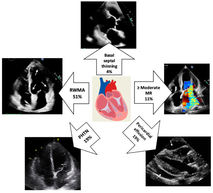
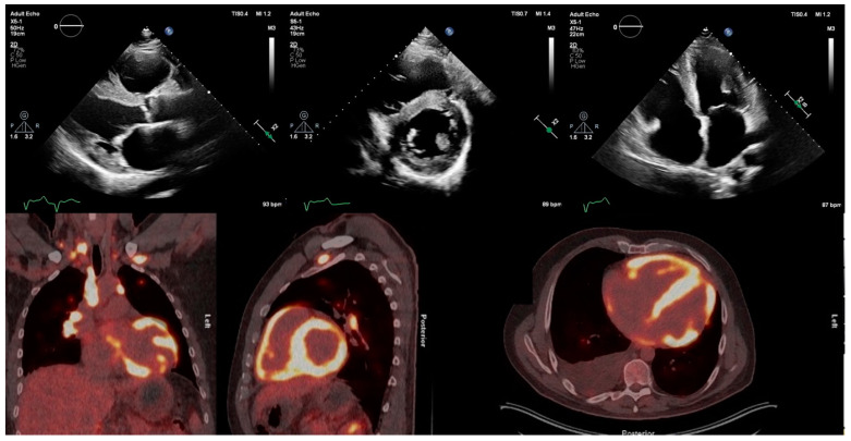
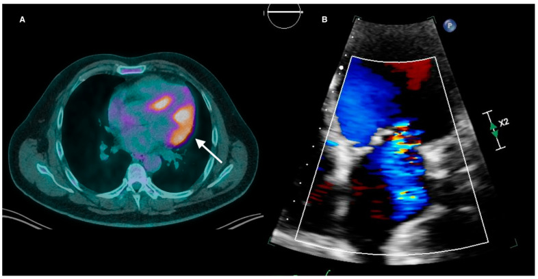
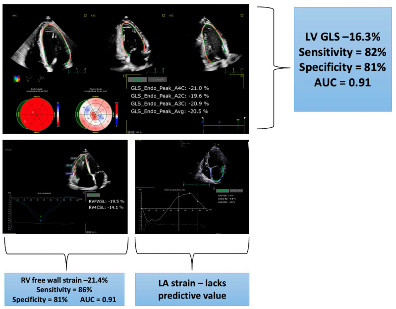
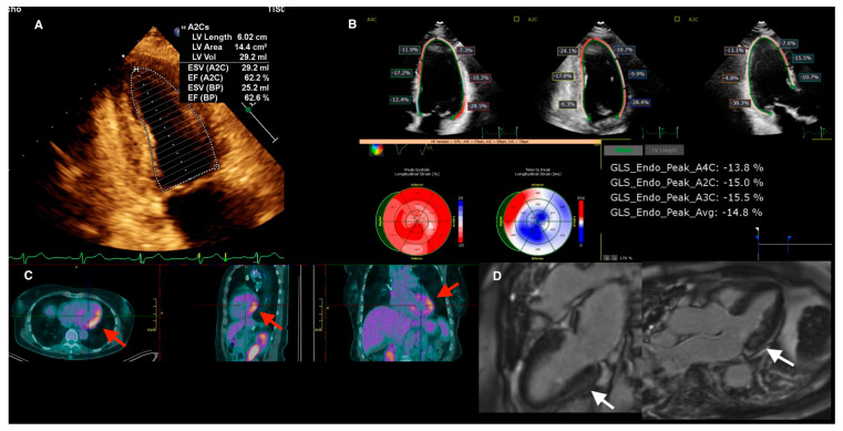
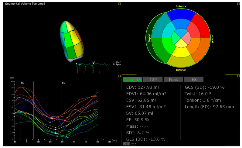
Cardiac sarcoidosis (CS) is a rare inflammatory disorder characterised by the presence of non-caseating granulomas within the myocardium. Contemporary studies have revealed that 25-30% of patients with systemic sarcoidosis have cardiac involvement, with detection rates increasing in the era of advanced cardiac imaging. The use of late gadolinium enhancement cardiac magnetic resonance and 18fluorodeoxy glucose positron emission tomography (FDG-PET) imaging has superseded endomyocardial biopsy for the diagnosis of CS. Echocardiography has historically been used as a screening tool with abnormalities triggering the need for advanced imaging, and as a tool to assess cardiac function. Regional wall thinning or aneurysm formation in a noncoronary distribution may indicate granuloma infiltration. Thinning of the basal septum in the setting of extracardiac sarcoidosis carries a high specificity for cardiac involvement. Abnormal myocardial echotexture and eccentric hypertrophy may be suggestive of active myocardial inflammation. The presence of right-ventricular involvement as indicated by free-wall aneurysms can mimic arrhythmogenic right-ventricular cardiomyopathy. More recently, the use of myocardial strain has increased the sensitivity of echocardiography in diagnosing cardiac involvement. Echocardiography is limited in prognostication, with impaired left-ventricular (LV) ejection fraction and LV dilatation being the only established independent predictors of mortality. More research is required to explore how advanced echocardiographic technologies can increase both the diagnostic sensitivity and prognostic ability of this modality in CS.
Keywords: FDG-PET; cardiac imaging; cardiac magnetic resonance; cardiac sarcoidosis; echocardiography; nuclear cardiology; sarcoid.
Conflict of interest statement
The other authors declare no conflicts of interest.
Figures
Similar articles
-
[Imaging diagnostics of cardiac sarcoidosis].Herz. 2023 Oct;48(5):366-371. doi: 10.1007/s00059-023-05208-z. Epub 2023 Sep 8. Herz. 2023. PMID: 37682338 Review. German.
-
Magnetic resonance imaging (MRI) for the assessment of myocardial viability: an evidence-based analysis.Ont Health Technol Assess Ser. 2010;10(15):1-45. Epub 2010 Jul 1. Ont Health Technol Assess Ser. 2010. PMID: 23074392 Free PMC article.
-
Isolated cardiac sarcoidosis diagnosed by electroanatomic voltage mapping-guided endomyocardial biopsy combined with magnetic resonance imaging and positron emission tomography.J Cardiol Cases. 2016 Jul 11;14(4):107-110. doi: 10.1016/j.jccase.2016.06.002. eCollection 2016 Oct. J Cardiol Cases. 2016. PMID: 30524562 Free PMC article.
-
Positron emission tomography for the assessment of myocardial viability: an evidence-based analysis.Ont Health Technol Assess Ser. 2005;5(16):1-167. Epub 2005 Oct 1. Ont Health Technol Assess Ser. 2005. PMID: 23074467 Free PMC article.
-
Current State and Future Directions of Multimodality Imaging in Cardiac Sarcoidosis.Front Cardiovasc Med. 2022 Jan 27;8:785279. doi: 10.3389/fcvm.2021.785279. eCollection 2021. Front Cardiovasc Med. 2022. PMID: 35155601 Free PMC article. Review.
Cited by
-
Contemporary Diagnostics of Cardiac Sarcoidosis: The Importance of Multimodality Imaging.Diagnostics (Basel). 2024 Aug 26;14(17):1865. doi: 10.3390/diagnostics14171865. Diagnostics (Basel). 2024. PMID: 39272650 Free PMC article. Review.
-
Arrhythmias in Cardiac Sarcoidosis: Management and Prognostic Implications.J Clin Med. 2024 May 28;13(11):3165. doi: 10.3390/jcm13113165. J Clin Med. 2024. PMID: 38892878 Free PMC article. Review.
-
Diagnostic Value of Comprehensive Echocardiographic Assessment Including Speckle-Tracking in Patients with Sarcoidosis Versus Healthy Controls: A Systematic Review and Meta-Analysis.Diagnostics (Basel). 2025 Mar 12;15(6):708. doi: 10.3390/diagnostics15060708. Diagnostics (Basel). 2025. PMID: 40150051 Free PMC article. Review.
-
Prognostic value of multimodality imaging in the contemporary management of cardiac sarcoidosis.Open Heart. 2024 Oct 27;11(2):e002989. doi: 10.1136/openhrt-2024-002989. Open Heart. 2024. PMID: 39462525 Free PMC article.
References
-
- Chiu C.-Z., Nakatani S., Zhang G., Tachibana T., Ohmori F., Yamagishi M., Kitakaze M., Tomoike H., Miyatake K. Prevention of left ventricular remodeling by long-term corticosteroid therapy in patients with cardiac sarcoidosis. Am. J. Cardiol. 2005;95:143–146. doi: 10.1016/j.amjcard.2004.08.083. - DOI - PubMed
-
- Kandolin R., Lehtonen J., Airaksinen J., Vihinen T., Miettinen H., Ylitalo K., Kaikkonen K., Tuohinen S., Haataja P., Kerola T., et al. Cardiac sarcoidosis: Epidemiology, characteristics, and outcome over 25 years in a nationwide study. Circulation. 2015;131:624–632. doi: 10.1161/CIRCULATIONAHA.114.011522. - DOI - PubMed
Publication types
LinkOut - more resources
Full Text Sources
