Review
. 2023 Aug 16;16(16):5642.
doi: 10.3390/ma16165642.

Recent Progress in Photonic Upconversion Materials for Organic Lanthanide Complexes

Affiliations
Review

Recent Progress in Photonic Upconversion Materials for Organic Lanthanide Complexes

Hong-Ju Yin et al. Materials (Basel). .

Abstract

Organic lanthanide complexes have garnered significant attention in various fields due to their intriguing energy transfer mechanism, enabling the upconversion (UC) of two or more low-energy photons into high-energy photons. In comparison to lanthanide-doped inorganic nanoparticles, organic UC complexes hold great promise for biological delivery applications due to their advantageous properties of controllable size and composition. This review aims to provide a summary of the fundamental concept and recent developments of organic lanthanide-based UC materials based on different mechanisms. Furthermore, we also detail recent applications in the fields of bioimaging and solar cells. The developments and forthcoming challenges in organic lanthanide-based UC offer readers valuable insights and opportunities to engage in further research endeavors.

Keywords: activator; mechanism; organic lanthanide complexes; sensitizer; upconversion luminescence.


Conflict of interest statement

The authors declare that the research was conducted in the absence of any commercial or financial relationships that could be construed as potential conflict of interest.

Figures

Figure 1
Figure 1
Schematic linear upconversion mechanisms operating in isolated molecules: (a) ESA, (b) ETU, (c) CU, and (d) CL. S, sensitizer; A, activator; G, ground state; E1/E2, excited state; blue, linear excitation; green, energy transfers; red, upconverted emission [32].
Scheme 1
Scheme 1
Diagram of ligand structures in organic complexes of four different mechanisms.
Figure 2
Figure 2
Diagram of organic complexes structures based on the ESA mechanism.
Figure 3
Figure 3
Diagram of organic complexes structures based on the ETU mechanism. (a), the structure diagram of the trinuclear complex [CrErCr(dipy-L4)3]9+ [20]; (b), the structure diagram of [CrEr(py-L4)3]6+ [21]; (c), the structure diagram of [(IR-806-L4)Er(L7)3]+ [37]; (d), the structure diagram of [Er(L6)3] [42]; (e), the structure diagram of [IR-806][Er(L5)4] [18]; (f), the structure diagram of {[Er(L2)]2F}+ [22]; (g), the structure diagram of the discrete bimetallic [Yb2Er]+ and the UCL intensity (545 nm) of [Yb2Er]+ in CH2Cl2 dependent on the quantity of [Bu4N]F under 2 Wcm−2 (left) and dependent on the quantity of [Bu4N]F under 2 Wcm−2 (right) [45]; (h), the structure diagram of [Er(L7)4(L8)2]+ and [Yb(L8)4)]3+ and the emission spectrum of polymer films [46]).
Figure 4
Figure 4
Diagram of organic complexes structures based on the CU mechanism. (“*” represents a species in an excited state; (a), the structure diagram of {[Yb(L9)]2Tb and the emission spectrum at 980 nm excitation [48]; (b), the structure diagram of {[Yb(L10)]2Tb} and the luminous mechanism [43]; (c), the structure diagram of [Yb8Tb(L11)16(OH)10]+ and the luminous mechanism [49]; (d), the structure diagram of [Yb(L1)3][Cr(L12)2] and the luminous mechanism [50]).
Figure 5
Figure 5
Diagram of organic complexes structures based on the CL mechanism. (“*” represents a species in an excited state; “**” represents a species in a virtual excited state; (a), the structure diagram of [Yb(L13)(L5)3] and the emission spectrum at the 950 nm excitation and the luminous mechanism [51]; (b), the structure diagram of [Yb9L7(OX)10]+ and the emission spectrum at 980 nm excitation and the luminous mechanism [19]).
Figure 6
Figure 6
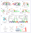
(a) Molecular structure, emission spectrum, and two-photon (λex = 860 nm) excitation-induced in vitro imaging in HeLa cells (40× magnification, λem = 500−800 nm) of the porphyrin-based Yb2 complex [67]. (b) The structures of the Sm and Yb complexes used for bioimaging experiments. Visible and NIR microscopy images of T24 cells fixed with PFA under laser irradiation at λex = 745 nm by using the [SmL15] emission in the visible channel and the NIR channel (the first row); for comparison, the [YbL16] emission in the NIR channel is also given (bottom right corner). Average intensities and associated standard deviations (bars) measured in central regions of interest (ROIs) of cells (bottom left corner) [69]. (c) The structure of the [YbL17]Otf used for bioimaging experiments. 2P−imaging of PFA−fixed T24 cells stained with [YbL17]Otf (c = 10−5 mol·L−1, 2P excitation at λex = 745 nm) using the custom-made NIR-to-NIR microscope. Detection in visible channel (<740 nm) and the NIR spectral (>840 nm) [70]. (d) MCLSFM images of the HeLa and H460 cells treated with complexes [Yb(L14)(L5)3] (the first row) and [Yb(L13)(L5)3] (the second row). NIR-to-visible cooperative upconversion emission shows nuclear and cytosolic localization of the complexes. λex = 950 nm, and λem = 490 nm [51].
Figure 7
Figure 7
(a) Chemical structures of Rubrene and Y6. (b) The UC mechanism proposed in this study with bilayer films of sensitizer and emitter. (c) UC emission from the rubrene/Y6 bilayer (orange) and pristine rubrene (grey) films irradiated by an 850-nm single-color LED at a power density of 101 mW·cm−2. For clarity, the PL intensity of the pure rubrene film was magnified by a factor of 100. (d) UC emission spectra of a rubrene/ITIC-Cl bilayer photovoltaic device under an applied bias with incident light from a 750-nm single-color LED with a power density of 78.4 mW·cm−2. (e) Photographs of UC emission by star-patterned NIR LED irradiation (750 nm, 71.7 mW·cm−2) on a flexible thin film [93].

References

    1. Liu S., Yan L., Huang J., Zhang Q., Zhou B. Controlling upconversion in emerging multilayer core–shell nanostructures: From fundamentals to frontier applications. Chem. Soc. Rev. 2022;51:1729–1765. doi: 10.1039/D1CS00753J. - DOI - PubMed
    1. Zheng X., Kankala R.K., Liu C.-G., Wang S.-B., Chen A.-Z., Zhang Y. Lanthanides-doped near-infrared active upconversion nanocrystals: Upconversion mechanisms and synthesis. Coord. Chem. Rev. 2021;438:213870–213887. doi: 10.1016/j.ccr.2021.213870. - DOI
    1. Healy C., Hermanspahn L., Kruger P.E. Photon upconversion in self-assembled materials. Coord. Chem. Rev. 2021;432:213756–213768. doi: 10.1016/j.ccr.2020.213756. - DOI
    1. Kwon O.S., Song H.S., Conde J., Kim H.-i., Artzi N., Kim J.-H. Dual-color emissive upconversion nanocapsules for differential cancer bioimaging In vivo. ACS Nano. 2016;10:1512–1521. doi: 10.1021/acsnano.5b07075. - DOI - PubMed
    1. Huang L., Le T., Huang K., Han G. Enzymatic enhancing of triplet–triplet annihilation upconversion by breaking oxygen quenching for background-free biological sensing. Nat. Commun. 2021;12:1898–1907. doi: 10.1038/s41467-021-22282-1. - DOI - PMC - PubMed

LinkOut - more resources