

Mechanical Properties and Liquid Absorption of Calcium Phosphate Composite Cements
- PMID: 37629944
- PMCID: PMC10456573
- DOI: 10.3390/ma16165653
Mechanical Properties and Liquid Absorption of Calcium Phosphate Composite Cements
Abstract
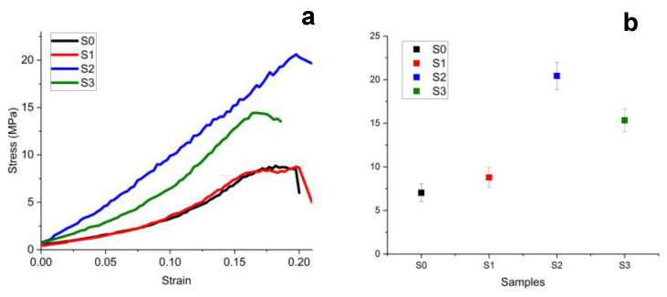
Calcium phosphate cements present increased biocompatibility due to their chemical composition being similar to that of the hydroxyapatite in the hard tissues of the living body. It has certain limitations due to its poor mechanical properties, such as low tensile strength and increased brittleness. Thus, the optimal way to improve properties is through the design of novel composite cements. The purpose was fulfilled using a 25% hydroxyethyl methacrylate (HEMA) mixed with 3% urethane dimethacrzlate (UDMA) base matrix with various ratios of polyethylene glycol (PEG 400) and polycaprolactone (PCL). Mineral filler is based on tricalcium phosphate (TCP) with different chitosan ratio used as bio-response enhancer additive. Four mixtures were prepared: S0-unfilled polymer matrix; S1 with 50% TCP filler; S2 with 50% chitosan + TCP filler; and S3 with 17.5% chitosan + TCP mixed with 17.5% nano hydroxyapatite (HA). The mechanical properties testing revealed that the best compressive strength was obtained by S2, followed by S3, and the worst value was obtained for the unfilled matrix. The same tendency was observed for tensile and flexural strength. These results show that the novel filler system increases the mechanical resistance of the TCP composite cements. Liquid exposure investigation reveals a relative constant solubility of the used filler systems during 21 days of exposure: the most soluble fillers being S3 and S2 revealing that the additivated TCP is more soluble than without additives ones. Thus, the filler embedding mode into the polymer matrix plays a key role in the liquid absorption. It was observed that additive filler enhances the hydrophobicity of UDMA monomer, with the matrix resulting in the lowest liquid absorption values, while the non-additivated samples are more absorbent due to the prevalence of hydrolytic aliphatic groups within PEG 400. The higher liquid absorption was obtained on the first day of immersion, and it progressively decreased with exposure time due to the relative swelling of the surface microstructural features. The obtained results are confirmed by the microstructural changes monitored by SEM microscopy. S3 and S2 present a very uniform and compact filler distribution, while S1 presents local clustering of the TCP powder at the contact with the polymer matrix. The liquid exposure revealed significant pore formation in S0 and S1 samples, while S3 and S2 proved to be more resistant against superficial erosion, proving the best resistance against liquid penetration.
Keywords: calcium phosphate; chitosan; composite cements; mechanical properties.
Conflict of interest statement
The authors declare no conflict of interest.
Figures


References
-
- Koons G.L., Diba M., Mikos A.G. Materials design for bone-tissue engineering. Nat. Rev. Mater. 2020;5:584–603. doi: 10.1038/s41578-020-0204-2. - DOI
LinkOut - more resources
Full Text Sources
