. 2023 Aug 18;14(8):1632.
doi: 10.3390/mi14081632.

Simultaneous Hydrostatic and Compressive Loading System for Mimicking the Mechanical Environment of Living Cartilage Tissue

Affiliations

Simultaneous Hydrostatic and Compressive Loading System for Mimicking the Mechanical Environment of Living Cartilage Tissue

Minki Chang et al. Micromachines (Basel). .

Abstract

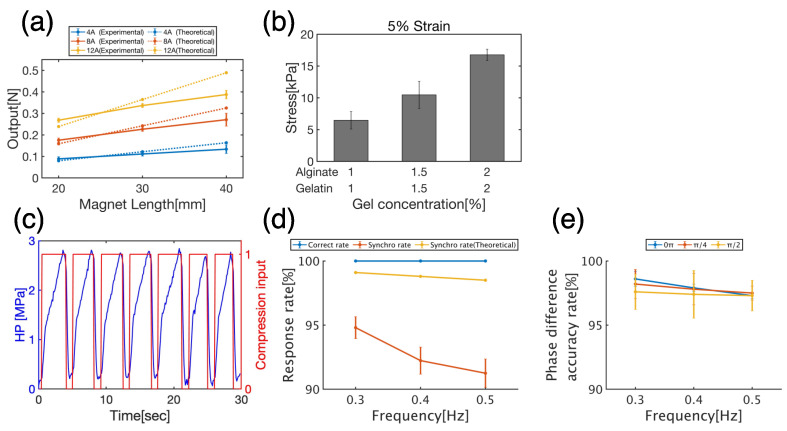
In vivo, articular cartilage tissue is surrounded by a cartilage membrane, and hydrostatic pressure (HP) and compressive strain increase simultaneously with the compressive stress. However, it has been impossible to investigate the effects of simultaneous loading in vitro. In this study, a bioreactor capable of applying compressive stress under HP was developed to reproduce ex vivo the same physical loading environment found in cartilage. First, a HP stimulation unit was constructed to apply a cyclic HP pressure-resistant chamber by controlling a pump and valve. A compression-loading mechanism that can apply compressive stress using an electromagnetic force was implemented in the chamber. The synchronization between the compression and HP units was evaluated, and the stimulation parameters were quantitatively evaluated. Physiological HP and compressive strain were applied to the chondrocytes encapsulated in alginate and gelatin gels after applying high HP at 25 MPa, which induced damage to the chondrocytes. It was found that compressive stimulation increased the expression of genes related to osteoarthritis. Furthermore, the simultaneous application of compressive strain and HP, which is similar to the physiological environment in cartilage, had an inhibitory effect on the expression of genes related to osteoarthritis. HP alone also suppressed the expression of osteoarthritis-related genes. Therefore, the simultaneous hydrostatic and compressive stress-loading device developed to simulate the mechanical environment in vivo may be an important tool for elucidating the mechanisms of disease onset and homeostasis in cartilage.

Keywords: articular cartilage; bioreactor; mechanical stimulation.


Conflict of interest statement

The authors declare no conflict of interest.

Figures

Figure 1
Figure 1
Schematic of the coil parameters. Parameters required to calculate theoretical values of the solenoid coil. “a1”: Inner radius (m), “a2”: Outer radius (m), “l”: Coil length (m), “x”: Distance (m) from “0”.
Figure 2
Figure 2
Schematic of the experimental flow. The samples were prepared using monolayer cells and gel scaffolds and incubated in an incubator for 24 h. After overstimulation of the samples with HHP at 25 MPa for 1 h to induce damage, the samples were loaded with physiological stimuli such as CHP (3 MPa, 0.3 Hz) and compression (3% strain). All cultures were conducted at 37 °C.
Figure 3
Figure 3
Designed bioreactor overview. (a) Schematic representation of the bioreactor. The HP unit is composed of water bath 1 (for water circulation in chambers), water bath 2 (for maintaining chamber temperature), a pump, chambers, an HP sensor, and a valve. The compression unit was composed of a solenoid coil, compression parts, and a control unit. (b) 3D-CAD modeling of compression parts. The upper panel shows the compression parts set inside the syringe. The transparent and black areas indicate the syringe rod and gasket, respectively. The lower figure shows the compression components. The yellow part indicates the indenter. A–E indicate the structures with the indenter. The green area represents the middle part. F–H indicate the structures with the middle part. The purple portion represents the sample holder. I–K indicate the structures with the sample holder. The red part represents the magnet holder. L–N indicate the structures with the magnet holder. (c) Images of compression parts. (c-1c-3) Images of the fabricated indenter. (c-4c-6) Images of the fabricated middle part. (c-7c-9) Images of the fabricated sample holder. (c-10c-12) Images of the fabricated magnet holder. (c-13,c-14) Images of the fabricated cover. (c-15,c-16) Images of the combination of compression components. (c-17,c-18) Images of the compression parts and samples set inside the syringe with culture medium.
Figure 4
Figure 4
Schematic of the compression control unit. (a) Electronic circuit diagram. The signal output from the HP unit is processed by the Arduino, leading to the control of the relay circuits through the pins 10, 11 for signal output and solenoid coil. (b) Relay circuit mechanism. When the electric current flows from 1 to 2, the electromagnetic force attracts the variable circuit and changes the electric current flow from 3 to 4 to that from 3 to 5. (c) Signal processing algorithm for synchronizing the compression unit to the HP unit.
Figure 5
Figure 5
Evaluation data for bioreactor and gel scaffold. (a) Theoretical and experimental compressive force outputs for each magnet length and current value. The graph shows the mean ± S.E. of three independent experiments. (b) Mechanical properties of scaffolds at each gel concentration. (c) Synchronization of HP (HP, blue line) and compressive input (red line). (d) Response rate, indicating the synchronization of the compression signal to HP. (e) Phase difference accuracy rate, indicating the accuracy with respect to the set phase difference of the compression signal. The graphs (d,e) show the mean ± S.E. of the results measured in 10 independent experiments with 30 cycles per experiment.
Figure 6
Figure 6
(a) Representative images of Calcein-AM/PI staining (scale bar = 100 µm) at 0 h. Green dots indicate live cells and red dots indicate dead cells. (b) Cell viability rate in control, CHP, Comp, and CHP+Comp groups (n = 4, mean ± S.E.).
Figure 7
Figure 7
Real-time PCR analysis. Gene expression of Socs3 in ATDC5 cells at control, CHP, Comp, and CHP+Comp (n = 4, mean ± S.E.). Gene expression was normalized to that of Rpl13a and the control. Asterisk indicates statistically significant difference (* p < 0.05).

Similar articles

References

    1. Mostakhdemin M., Nand A., Ramezani M. Articular and Artificial Cartilage, Characteristics, Properties and Testing Approaches—A Review. Polymers. 2021;13:2000. doi: 10.3390/polym13122000. - DOI - PMC - PubMed
    1. Wong M., Carter D.R. Articular cartilage functional histomorphology and mechanobiology: A research perspective. Bone. 2003;33:1–13. doi: 10.1016/S8756-3282(03)00083-8. - DOI - PubMed
    1. Urban J.P.G. The chondrocyte: A cell under pressure. Rheumatology. 1994;33:901–908. doi: 10.1093/rheumatology/33.10.901. - DOI - PubMed
    1. Lu X.L., Mow V.C. Biomechanics of articular cartilage and determination of material properties. Med. Sci. Sports Exerc. 2008;40:193–199. doi: 10.1249/mss.0b013e31815cb1fc. - DOI - PubMed
    1. Elder B.D., Athanasiou K.A. HP in articular cartilage tissue engineering: From chondrocytes to tissue regeneration. Tissue Eng. Part B Rev. 2009;15:43–53. doi: 10.1089/ten.teb.2008.0435. - DOI - PMC - PubMed