. 2023 Aug 16;28(16):6080.
doi: 10.3390/molecules28166080.

Binding Specificity of a Novel Cyclo/Maltodextrin-Binding Protein and Its Role in the Cyclodextrin ABC Importer System from Thermoanaerobacterales

Affiliations

Binding Specificity of a Novel Cyclo/Maltodextrin-Binding Protein and Its Role in the Cyclodextrin ABC Importer System from Thermoanaerobacterales

Jorge Aranda-Caraballo et al. Molecules. .

Abstract

Extracellular synthesis of functional cyclodextrins (CDs) as intermediates of starch assimilation is a convenient microbial adaptation to sequester substrates, increase the half-life of the carbon source, carry bioactive compounds, and alleviate chemical toxicity through the formation of CD-guest complexes. Bacteria encoding the four steps of the carbohydrate metabolism pathway via cyclodextrins (CM-CD) actively internalize CDs across the microbial membrane via a putative type I ATP-dependent ABC sugar importer system, MdxEFG-(X/MsmX). While the first step of the CM-CD pathway encompasses extracellular starch-active cyclomaltodextrin glucanotransferases (CGTases) to synthesize linear dextrins and CDs, it is the ABC importer system in the second step that is the critical factor in determining which molecules from the CGTase activity will be internalized by the cell. Here, structure-function relationship studies of the cyclo⁄maltodextrin-binding protein MdxE of the MdxEFG-MsmX importer system from Thermoanaerobacter mathranii subsp. mathranii A3 are presented. Calorimetric and fluorescence studies of recombinant MdxE using linear dextrins and CDs showed that although MdxE binds linear dextrins and CDs with high affinity, the open-to-closed conformational change is solely observed after α- and β-CD binding, suggesting that the CM-CD pathway from Thermoanaerobacterales is exclusive for cellular internalization of these molecules. Structural analysis of MdxE coupled with docking simulations showed an overall architecture typically found in sugar-binding proteins (SBPs) that comprised two N- and C-domains linked by three small hinge regions, including the conserved aromatic triad Tyr193/Trp269/Trp378 in the C-domain and Phe87 in the N-domain involved in CD recognition and stabilization. Structural bioinformatic analysis of the entire MdxFG-MsmX importer system provided further insights into the binding, internalization, and delivery mechanisms of CDs. Hence, while the MdxE-CD complex couples to the permease subunits MdxFG to deliver the CD into the transmembrane channel, the dimerization of the cytoplasmatic promiscuous ATPase MsmX triggers active transport into the cytoplasm. This research provides the first results on a novel thermofunctional SBP and its role in the internalization of CDs in extremely thermophilic bacteria.

Keywords: CM-CD; MdxE; SBP; carbohydrate metabolism; starch-converting pathway.


Conflict of interest statement

The authors declare no conflict of interest.

Figures

Figure 1
Figure 1
Purification of recombinant MdxE. (A) SEC-DLS coupled experiment of MdxE. Right inset: molecular weight (Mw), polydispersity index (Mw/Mn), and weight fraction (Wt Fr) of chromatographic peaks 1 and 2. Note that the MdxE aggregates (4021 kDa, peak 1) correspond to 28.6% of the total injected sample, showing the tendency of MdxE to form aggregates. (B) Coomassie Blue-stained SDS-PAGE gel (12%). Lane 1, molecular-weight markers (Bio-Rad, labeled in kDa). Lane 2, insoluble fraction of MdxE production. Lane 3, soluble fraction of MdxE production after a heat treatment procedure (60 °C, 20 min). Lane 4, purified MdxE after Ni2+-affinity chromatography. Lane 5, purified recombinant MdxE with optimal monodispersity (Mw/Mn = 1.04, peak 2) after SEC-DLS analysis.
Figure 2
Figure 2
(A) Homology model of MdxE showing the N–domain (blue), C–domain (red), and hinge regions (green) in two views related by a horizontal rotation of 90 degrees. The residues comprising the N–domain, C–domain, and hinge regions are indicated below the MdxE model, following the same color code. The conserved aromatic triad involved in sugar recognition in the C–domain (red molecular surface) and the hydrophobic residue in the N–domain (blue molecular surface) involved in obtaining the closed form are shown in (B) MdxE (black cylinders), (C) TvuCMBP (cyan cylinders) in complex with β–CD (PDB ID: 2ZYN, [36], and (D) EcoMBP (yellow cylinders) in complex with G2 (PDB ID: 1ANF) [37,38,39].
Figure 3
Figure 3
ITC studies of MdxE titrated with (A) α−CD, (B) β−CD, (C) γ−CD, (D) maltose (G2), (E) maltotriose (G3), (F) maltopentaose (G5), (G) maltohexaose (G6), and (H) maltoheptaose (G7).
Figure 4
Figure 4
Fluorescence determination of the open-to-closed conformational change by MdxE complexed with CDs and linear dextrins. Note that the scans were taken in the 400–600 nm range. (A) MdxE-ligand complexes (16 μM) bound to ANS. (B) Ligands with ANS (blank). (C) The increasing intensity was monitored by relative fluorescence unit (RFU) measurements at λmax = 485 nm as a function of MdxE-ligand concentration (2–14 μM). Note that each Ho value is obtained as the slope of each of the linear functions [47].
Figure 5
Figure 5
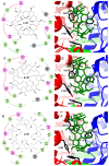
Docked structures of α-, β-, and γ-CDs in the sugar-binding site of MdxE. (A,B) MdxE/α-CD. (C,D) MdxE/β-CD. (E,F) MdxE/γ-CD. The two-dimensional (2D) interaction plots show the hydrogen bonds between glucose (G1) and the side chain of a residue in green, hydrophobic interactions in violet, and hydrogen bonds with the main chain atoms in gray. The 3D docked structures exhibit the key residues (black cylinders) in the N-domain (blue) and C-domain (red) involved in CD recognition. The residues in the 2D interaction plots are linked with red, blue, and green lines representing the C-domain, N-domain, and hinge regions, respectively. Note that the absence of Glu155 from hinge region I in the MdxE/γ-CD complex might affect obtaining the closed form. Distances are in Å.
Figure 6
Figure 6
Amino acid sequence alignment of EcoMBP, TvuCMBP, and MdxE. Note the conserved Glu residue (black triangle) in hinge region I of SBPs. Sequence alignment was performed using ClustalW [49].
Figure 7
Figure 7
Homology model of the entire type I ABC importer system, MdxEFG-MsmX, from T. mathranii subsp. mathranii. (A) Close-up of the binding site showing MdxE on the magenta molecular surface, the spoon loop (P3) of MdxG in goldenrod ribbon representation, and α-CD in black and red cylinders. Note that Gln256 from MdxG seems to be involved in removing the CDs from the MdxE-binding site. (B) Close-up of the binding pocket showing MdxF and MdxG in green and goldenrod ribbon representations, respectively, the aromatic triad Trp119/Phe177/Tyr230 in green cylinders, and α-CD in black and red cylinders. (C) Close-up of CH from MdxF (green) showing the conserved Glu190 in green and red cylinders, directly interacting with Arg48 in gray and blue cylinders from the Walker A (green) motif in MsmX. The Walker B motif is shown in deep blue. The NBD subunit is shown on a gray molecular surface. (D) Close-up of the detailed interactions at the active side of the NBD with a docked ATP molecule. The MsmX subunit is shown in gray ribbon representation, as well as signature motifs LSGGQ (red), Walker A (dark green), and D-loop (orange), which along with the Walker B motif, encompass most of the residues (Tyr13, Lys43, Glu160, and His193) needed for ATPase activity and signal transmission between the TMD-NBD. Note that the interaction between Lys43 and the coordinated Mg2+ atom (gray sphere) is crucial for the cleavage of γ-phosphate from the ATP molecule.
Figure 8
Figure 8
Proposed internalization mechanism of the type I ABC importer system, MdxEFG-MsmX, from Thermoanaerobacterales. The mechanism encompasses five main steps: (i) Ligand recognition by MdxE. (ii) Coupling of MdxE to the MdxFG-MsmX core unit and transition into a PTS. (iii) Ligand transfer into the MdxFG transmembrane channel via an OF conformation obtained by MsmX dimerization. (iv) The dimerization process is completed, and ATP hydrolysis occurs, releasing MdxE from the core unit, ADP and Pi. (v) The ligand is finally translocated into the cytoplasm, allowing transition into an IF conformation for the next MdxE cycle. The three states of MdxEFG-MsmX from Thermoanaerobacterales were built upon MalEFG-MalK structure from E. coli (PDB IDs: 3FH6, 3PUZ, and 3PUV) [50,51,55].

References

    1. Fiedler G., Pajatsch M., Böck A. Genetics of a Novel Starch Utilisation Pathway Present in Klebsiella oxytoca. J. Mol. Biol. 1996;256:279–291. doi: 10.1006/jmbi.1996.0085. - DOI - PubMed
    1. Shim J.-H., Park J.-T., Hong J.-S., Kim K.W., Kim M.-J., Auh J.-H., Kim Y.-W., Park C.-S., Boos W., Kim J.-W., et al. Role of Maltogenic Amylase and Pullulanase in Maltodextrin and Glycogen Metabolism of Bacillus subtilis 168. J. Bacteriol. 2009;191:4835–4844. doi: 10.1128/JB.00176-09. - DOI - PMC - PubMed
    1. Oslowski D.M., Jung J.-H., Seo D.-H., Park C.-S., Holden J.F. Production of Hydrogen from α-1,4- and β-1,4-Linked Saccharides by Marine Hyperthermophilic Archaea. Appl. Environ. Microbiol. 2011;77:3169–3173. doi: 10.1128/AEM.01366-10. - DOI - PMC - PubMed
    1. Grégorio C. A Review: A History of Cyclodextrins. Chem. Rev. 2014;114:10940–10975. - PubMed
    1. Uitdehaag J.C., van der Veen B.A., Dijkhuizen L., Dijkstra B.W. Catalytic mechanism and product specificity of cyclodextrin glycosyltransferase, a prototypical transglycosylase from the α-amylase family. Enzym. Microb. Technol. 2002;30:295–304. doi: 10.1016/S0141-0229(01)00498-7. - DOI

Supplementary concepts

LinkOut - more resources