. 2023 Aug 16;15(16):3596.
doi: 10.3390/nu15163596.

Anticancer Effect of Gallic Acid on Acidity-Induced Invasion of MCF7 Breast Cancer Cells

Affiliations

Anticancer Effect of Gallic Acid on Acidity-Induced Invasion of MCF7 Breast Cancer Cells

Ran Hong et al. Nutrients. .

Abstract

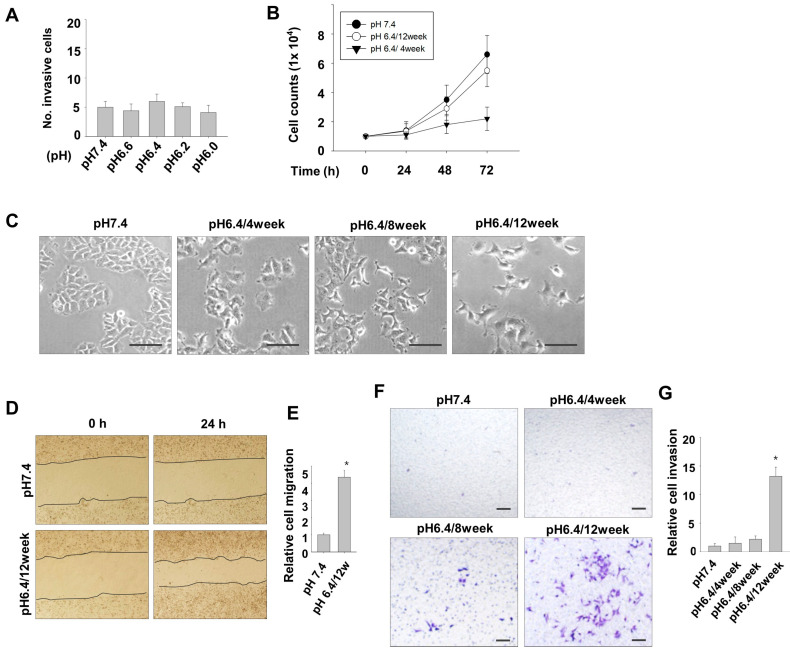
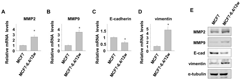
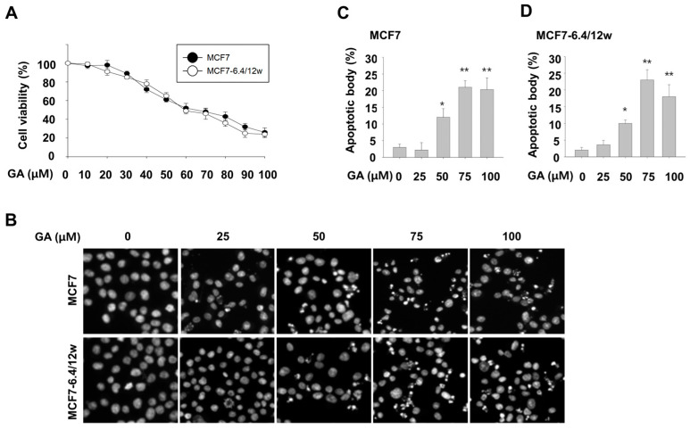
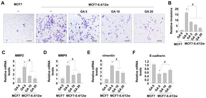
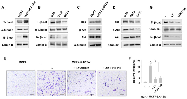
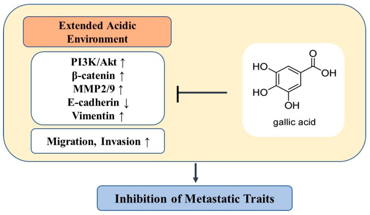
The acidic tumor environment has emerged as a crucial factor influencing the metastatic potential of cancer. We investigated the effect of an acidic environment on the acquisition of metastatic properties in MCF7 breast cancer cells and explored the inhibitory effects of gallic acid. Prolonged exposure to acidic culture conditions (over 12 weeks at pH 6.4) induced the acquisition of migratory and invasive properties in MCF7 cells, accompanied by increased expression of Matrix Metalloproteinase 2 and 9 (MMP2 and MMP9, respectively), together with alterations in E-cadherin, vimentin, and epithelial-to-mesenchymal transition markers. Gallic acid effectively inhibited the survival of acidity-adapted MCF7 (MCF7-6.4/12w) cells at high concentrations (>30 μM) and reduced metastatic characteristics induced by acidic conditions at low concentration ranges (5-20 μM). Moreover, gallic acid suppressed the PI3K/Akt pathway and the nuclear accumulation of β-catenin, which were elevated in MCF7-6.4/12w cells. These findings highlight the potential of gallic acid as a promising therapeutic agent for metastatic traits in breast cancer cells under acidic conditions.

Keywords: MCF7 cells; acidic tumor environment; gallic acid.


Conflict of interest statement

The authors declare no conflict of interest.

Figures

Figure 1
Figure 1
Long-term exposure to environmental acidity induces migration and invasive potential in MCF7 cells. (A) MCF7 cells exposed to a normal pH medium (pH 7.4) and various acidic pH-adjusted culture media (pH 6.6, 6.4, 6.2, and 6.0) were analyzed for invasive activity. The numbers of invaded cells are depicted graphically. (B) Cell growth was assessed by culturing MCF7 cells from the normal pH (7.4) condition, pH 6.4 for 4 weeks, and pH 6.4 for 12 weeks in a 24-well plate with regular medium for 3 d. (C) Phase-contrast images were captured for cells cultured under normal pH condition and those cultured at pH 6.4, for 4, 8, and 12 weeks. (D,E) Cells were cultured in either pH 7.4 or 6.4 medium for 12 weeks, and cell migration was evaluated using a wound healing assay at 24 h after creating the scratch (D), and the relative migration distance is graphically represented (E). (F,G) Cells cultured at pH 7.4 condition and those cultured at pH 6.4, for 4, 8, and 12 weeks were analyzed for invasion activity using a Matrigel-coated Transwell plate. Invaded cells were detected under a microscope (F), and the relative number of invaded cells are graphically represented (G). * p < 0.05 vs. pH 7.4. Scale bar = 100 μm.
Figure 2
Figure 2
Long-term exposure to environmental acidity induces epithelial-to-mesenchymal transition (EMT)-like features. The mRNA expression of Matrix Metalloproteinases (MMP) 2, MMP9, E-cadherin, and vimentin was analyzed using real-time PCR (AD) and Western blot assay (E) in MCF7 cells and MCF7 cells cultured for over 12 weeks at pH 6.4 (MCF7-6.4/12w cells). * p < 0.05 vs. pH 7.4.
Figure 3
Figure 3
Gallic acid exerts inhibitory effect on the survival of both normal and acidity-adapted MCF7 cells. Cell viability was determined using the EZ-Cytox assay 48 h after treating MCF7 and MCF7-6.4/12w cells with gallic acid at specified concentrations (A). (BD) Following treatment with gallic acid at different concentrations, MCF7 and MCF7-6.4/12w cells were stained with Hoechst 33342 to visualize apoptotic bodies under a fluorescent microscope (B), and the number of apoptotic nuclei in MCF7 (C) and MCF7-6.4/12w cells (D) was quantified. * p < 0.05, ** p < 0.01 vs. pH 7.4.
Figure 4
Figure 4
Low concentrations of gallic acid decreases acidity-induced metastatic characteristics in MCF7-6.4/12w cells. (A,B) Following 48 h of treatment with 5, 10, and 20 μM of gallic acid in MCF7-6.4/12w cells, an invasion assay was conducted (A) and the relative number of invaded cells compared to the normal MCF7 cells (B). (CF) MCF7-6.4/12w cells were treated with 0, 10, and 20 μM of gallic acid for 48 h, and the mRNA expression levels of MMP2 (C), MMP9 (D), vimentin (E), and E-cadherin (F), and were analyzed using real-time PCR. The relative mRNA expression levels were calculated with the number of untreated normal MCF7 cells as the reference. * p < 0.05 vs. MCF7, # p < 0.05 vs. untreated MCF7-6.4/12w. Scale bar = 100 μm.
Figure 5
Figure 5
The inhibitory effect of gallic acid on the invasion of MCF7-6.4/12w cells is related to the inhibition of the PI3K/Akt signaling-controlled β-catenin pathway. (AD) Western blot assay was used to analyze total and nuclear β-catenin levels in MCF7 and MCF7-6.4/12w cells (A) and to assess the expression levels of p85, pAKT, and total AKT (C). In MCF7-6.4/12w cells, after treatment with 0, 10, and 20 μM of gallic acid, total and nuclear β-catenin levels (B), along with the expression levels of p85, pAKT, and total AKT (D), were also evaluated using a Western blot assay. (E,F) An invasion assay was conducted using normal MCF7 cells and MCF7-6.4/12w cells treated with either 1 μM LY294002, 1 μM AKT Inhibitor VIII, or untreated (E), and the relative ratio of invaded cells compared to normal MCF7 cells is represented (F). (G) MCF7-6.4/12w cells treated with 1 μM LY294002, 1 μM AKT Inhibitor VIII, or untreated, were analyzed by Western blot assay to detect total and nuclear β-catenin levels. Lamin B is nuclear loading control. * p < 0.05 vs. MCF7, # p < 0.05 vs. untreated MCF7-6.4/12w. Scale bar = 100 μm.
Figure 6
Figure 6
A schematic model illustrating the role of gallic acid in inhibiting metastatic traits induced by prolonged environmental acidity. ↑: upregulation, ↓: downregulation.

References

    1. Arnold M., Morgan E., Rumgay H., Mafra A., Singh D., Laversanne M., Vignat J., Gralow J.R., Cardoso F., Siesling S., et al. Current and future burden of breast cancer: Global statistics for 2020 and 2040. Breast. 2022;66:15–23. doi: 10.1016/j.breast.2022.08.010. - DOI - PMC - PubMed
    1. Nathanson S.D., Detmar M., Padera T.P., Yates L.R., Welch D.R., Beadnell T.C., Scheid A.D., Wrenn E.D., Cheung K. Mechanisms of breast cancer metastasis. Clin. Exp. Metastasis. 2022;39:117–137. doi: 10.1007/s10585-021-10090-2. - DOI - PMC - PubMed
    1. Christiansen J.J., Rajasekaran A.K. Reassessing epithelial to mesenchymal transition as a prerequisite for carcinoma invasion and metastasis. Cancer Res. 2006;66:8319–8326. doi: 10.1158/0008-5472.CAN-06-0410. - DOI - PubMed
    1. Neophytou C.M., Panagi M., Stylianopoulos T., Papageorgis P. The Role of Tumor Microenvironment in Cancer Metastasis: Molecular Mechanisms and Therapeutic Opportunities. Cancers. 2021;13:2053. doi: 10.3390/cancers13092053. - DOI - PMC - PubMed
    1. Vaupel P., Multhoff G. Revisiting the Warburg effect: Historical dogma versus current understanding. J. Physiol. 2021;599:1745–1757. doi: 10.1113/JP278810. - DOI - PubMed