Review
. 2023 Aug 10;15(8):2118.
doi: 10.3390/pharmaceutics15082118.

Biopolymers and Their Application in Bioprinting Processes for Dental Tissue Engineering

Affiliations
Review

Biopolymers and Their Application in Bioprinting Processes for Dental Tissue Engineering

Suhon Kim et al. Pharmaceutics. .

Abstract

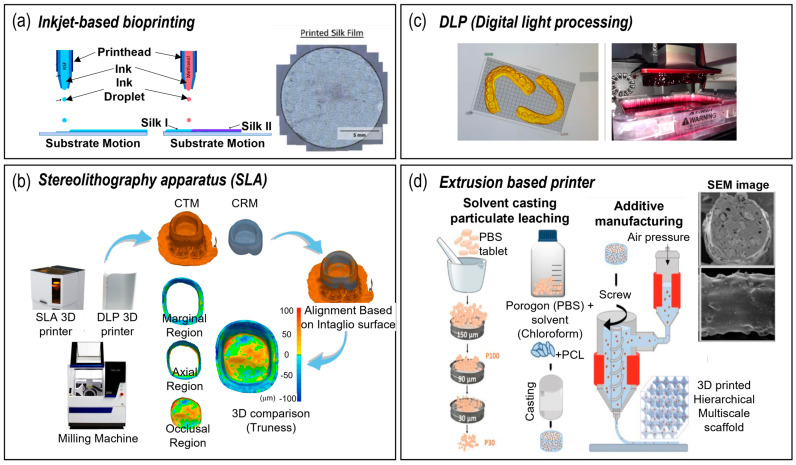
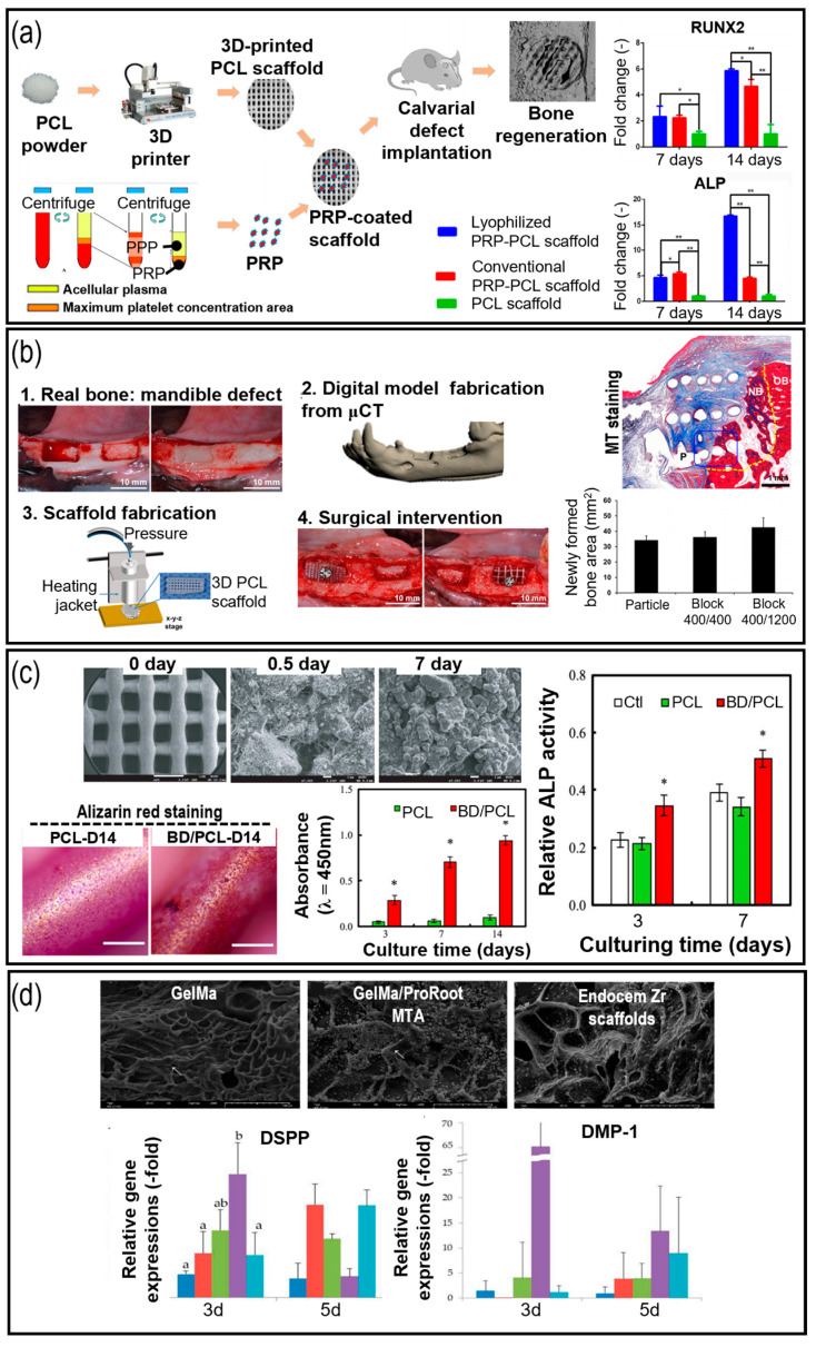
Dental tissues are composed of multiple tissues with complex organization, such as dentin, gingiva, periodontal ligament, and alveolar bone. These tissues have different mechanical and biological properties that are essential for their functions. Therefore, dental diseases and injuries pose significant challenges for restorative dentistry, as they require innovative strategies to regenerate damaged or missing dental tissues. Biomimetic bioconstructs that can effectively integrate with native tissues and restore their functionalities are desirable for dental tissue regeneration. However, fabricating such bioconstructs is challenging due to the diversity and complexity of dental tissues. This review provides a comprehensive overview of the recent developments in polymer-based tissue engineering and three-dimensional (3D) printing technologies for dental tissue regeneration. It also discusses the current state-of-the-art, focusing on key techniques, such as polymeric biomaterials and 3D printing with or without cells, used in tissue engineering for dental tissues. Moreover, the final section of this paper identifies the challenges and future directions of this promising research field.

Keywords: 3D printing; dental tissues; polymers; tissue engineering.


Conflict of interest statement

The authors declare no conflict of interest.

Figures

Figure 1
Figure 1
A schematic representation delineating the tissue engineering approach for the regeneration of periodontal tissues, which were obtained using 3D bioprinted scaffolds, diverse stem cells, and signaling molecules.
Figure 2
Figure 2
3D fabrication systems for dental tissue engineering. (a) Inkjet-based bioprinting [44]; (b) sterolithography [45]; (c) digital light processing [46]; and (d) extrusion-based fabrication method [47].
Figure 3
Figure 3
Application of polymeric materials used for dental tissue engineering. (a) Effects of platelet-rich plasma (PRP)-coated polycaprolactone (PCL) scaffold in osteogenesis of human dental stem cells (* p < 0.05 and ** p < 0.005) [93]; (b) implantation of β-TCP/PCL scaffold to alveolar defect in rats and Masson’s Trichrome (MT) staining results [95]; (c) SEM and ARS images of dentine incorporated PCL scaffold, cellular proliferation and alkaline phosphatase (ALP) activities of human dental pulp stem cells [96] (* p < 0.05); (d) incorporation of mineral trioxide aggregate to photo-crosslinkable methacrylated gelatin scaffolds (a, b, and ab, represents statistically significant differences (p < 0.05)) [97].
Figure 4
Figure 4
The application of cell-printing and cell-laden structures for dental tissue regeneration. (a) Application of bone decellularized extracellular matrix and β-tricalcium phosphate (β-TCP) for accelerated osteo/odontogenic differentiation of human dental pulp stem cells [112]; (b) incorporation of demineralized dentin matrix particles to fibronogen/gelatin/hyaluronic acid/glycerol to evoke efficient odontogenic differentiation [113]; (c) mechanically enhanced titanium/collagen hybrid structures for dental implants [115]; (d) incorporation of nano-sized demineralized human dentin matrix particles into alginate hydrogel for enhanced dental repair [116]; (e) top- and side-view photographic images of the Col bio-ink/SrCS bi-layer structure, the proliferation of encapsulated human gingiva fibroblasts (hGF), representative µCT images of osteoporotic rabbits’ cranial bone defect model after being implanted for 12 weeks, and µCT-quantified histograms of bone volume/total volume (BV/TV) and trabecular thickness (Tb.Th) (* indicates a significant difference (p < 0.05) compared to SrCS and # indicates a significant difference (p < 0.05) compared to bi-layer) [117].

Similar articles

Cited by

References

    1. Kim T.G., Shin H., Lim D.W. Biomimetic scaffolds for tissue engineering. Adv. Funct. Mater. 2012;22:2446–2468.
    1. Nakashima M., Reddi A.H. The application of bone morphogenetic proteins to dental tissue engineering. Nat. Biotechnol. 2003;21:1025–1032. doi: 10.1038/nbt864. - DOI - PubMed
    1. Galler K.M., D’Souza R.N., Hartgerink J.D. Biomaterials and their potential applications for dental tissue engineering. J. Mater. Chem. 2010;20:8730–8746.
    1. Jazayeri H.E., Lee S.-M., Kuhn L., Fahimipour F., Tahriri M., Tayebi L. Polymeric scaffolds for dental pulp tissue engineering: A review. Dent. Mater. 2020;36:e47–e58. - PubMed
    1. Tang G., Tan Z., Zeng W., Wang X., Shi C., Liu Y., He H., Chen R., Ye X. Recent advances of chitosan-based injectable hydrogels for bone and dental tissue regeneration. Front. Bioeng. Biotechnol. 2020;8:587658. doi: 10.3389/fbioe.2020.587658. - DOI - PMC - PubMed

LinkOut - more resources