. 2023 Jul 30;15(8):1665.
doi: 10.3390/v15081665.

Compounding Achromobacter Phages for Therapeutic Applications

Affiliations

Compounding Achromobacter Phages for Therapeutic Applications

Ana Georgina Cobián Güemes et al. Viruses. .

Abstract

Achromobacter species colonization of Cystic Fibrosis respiratory airways is an increasing concern. Two adult patients with Cystic Fibrosis colonized by Achromobacter xylosoxidans CF418 or Achromobacter ruhlandii CF116 experienced fatal exacerbations. Achromobacter spp. are naturally resistant to several antibiotics. Therefore, phages could be valuable as therapeutics for the control of Achromobacter. In this study, thirteen lytic phages were isolated and characterized at the morphological and genomic levels for potential future use in phage therapy. They are presented here as the Achromobacter Kumeyaay phage collection. Six distinct Achromobacter phage genome clusters were identified based on a comprehensive phylogenetic analysis of the Kumeyaay collection as well as the publicly available Achromobacter phages. The infectivity of all phages in the Kumeyaay collection was tested in 23 Achromobacter clinical isolates; 78% of these isolates were lysed by at least one phage. A cryptic prophage was induced in Achromobacter xylosoxidans CF418 when infected with some of the lytic phages. This prophage genome was characterized and is presented as Achromobacter phage CF418-P1. Prophage induction during lytic phage preparation for therapy interventions require further exploration. Large-scale production of phages and removal of endotoxins using an octanol-based procedure resulted in a phage concentrate of 1 × 10⁹ plaque-forming units per milliliter with an endotoxin concentration of 65 endotoxin units per milliliter, which is below the Food and Drugs Administration recommended maximum threshold for human administration. This study provides a comprehensive framework for the isolation, bioinformatic characterization, and safe production of phages to kill Achromobacter spp. in order to potentially manage Cystic Fibrosis (CF) pulmonary infections.

Keywords: Achromobacter phage; phage production; phage therapy; prophage induction.


Conflict of interest statement

The authors declare no conflict of interest.

Figures

Figure 1
Figure 1
Phage proteomic tree displaying the relationships of Achromobacter phages. Publicly available Achromobacter phages, including the Côte d’Ivoire collection (n = 16), were compared to the Kumeyaay phages isolated in this study (n = 13) and the induced prophage CF418-P1. The Phage Proteomic Tree was calculated using the Viral Proteomic Tree server (https://www.genome.jp/viptree/) (accessed on March 2022). Selected phages from the ViPTree database were used to compare clades.
Figure 2
Figure 2
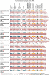
Genome comparisons of Achromobacter phages belonging to clade phiAxp1. All phages were aligned with the terL gene at the left end of the linear map. Genome maps were generated using the VIP server. Phages marked with a pink circle were isolated and described as part of our study.
Figure 3
Figure 3
Genome comparisons of the Achromobacter phages belonging to clade JWX. Phages were aligned with the terL gene at the left end of the linear map (generated by ViPtree). Genome maps were generated using the VIP server. Phages marked with a pink circle were isolated and described as part of our study.
Figure 4
Figure 4
Achromobacter prophage was induced when the host was infected with other lytic phages. (A) Prophage in A. xylosoxidans CF418. The prophage genome was assembled from phage lysates LB5, LB7, and LB8 using SPAdes. Prophage annotation using PATRIC, CDD, HMMER, and ANNs. (B) Identity of Achromobacter phage CF418-P1 to closest bacteria genomes. (C) Prophage in A. xylosoxidans CF418. Fragment recruitment plots of phage lysate against the Achromobacter reference genome. The prophage region recruits reads around 3.9 Mbp (highlighted in the red vertical rectangle).
Figure 5
Figure 5
Host range of the Kumeyaay and Côte d’Ivoire Achromophages. Achromobacter strains isolated from the sputum of CF patients and characterized by the UCSD Medical Center clinical diagnostic laboratory were used to test host ranges. The Achromobacter reference strain (HM-235) was also used for testing host ranges. Strain AC116, also known as CF116, is Achromobacter ruhlandii. The host range was tested by performing spot tests on a lawn of the host bacteria. Lysate LB5, LB7, and LB8 contain the prophage CF418-P1 (Supplementary Figure S3). NA, not available.
Figure 6
Figure 6
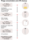
Protocol for Achromobacter phage nyaak high-titer lysate production and endotoxin removal. The Achromobacter phage nyaak was produced in Achromobacter ruhlandii CF116. Final endotoxin units were measured with the Endozyme II kit from Biomerieux.
Figure 7
Figure 7
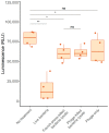
Cytotoxic effects of bacteria and/or phages on A549 cells. The Achromobacter phage nyaak and Achromobacter ruhlandii CF116 were used in the experiments. Ns = not significant, * p-value < 0.01, ** p-values < 0.001.

References

    1. Ridderberg W., Nielsen S.M., Nørskov-Lauritsen N. Genetic Adaptation of Achromobacter sp. during Persistence in the Lungs of Cystic Fibrosis Patients. PLoS ONE. 2015;10:e0136790. doi: 10.1371/journal.pone.0136790. - DOI - PMC - PubMed
    1. Jakobsen T.H., Hansen M.A., Jensen P.Ø., Hansen L., Riber L., Kolpen M., Hansen C.R., Rønne Hansen C., Ridderberg W., Eickhardt S., et al. Complete Genome Sequence of the Cystic Fibrosis Pathogen Achromobacter xylosoxidans NH44784-1996 Complies with Important Pathogenic Phenotypes. PLoS ONE. 2013;8:e68484. doi: 10.1371/journal.pone.0068484. - DOI - PMC - PubMed
    1. Tena D., González-Praetorius A., Pérez-Balsalobre M., Sancho O., Bisquert J. Urinary Tract Infection Due to Achromobacter xylosoxidans: Report of 9 Cases. Scand. J. Infect. Dis. 2008;40:84–87. doi: 10.1080/00365540701558714. - DOI - PubMed
    1. Tokuyasu H., Fukushima T., Nakazaki H., Shimizu E. Infective Endocarditis Caused by Achromobacter xylosoxidans: A Case Report and Review of the Literature. Intern. Med. 2012;51:1133–1138. doi: 10.2169/internalmedicine.51.6930. - DOI - PubMed
    1. Manckoundia P., Mazen E., Coste A.S., Somana S., Marilier S., Duez J.M., Camus A., Popitean L., Bador J., Pfitzenmeyer P. A Case of Meningitis Due to Achromobacter xylosoxidans Denitrificans 60 Years after a Cranial Trauma. Med. Sci. Monit. 2011;17:2010–2012. doi: 10.12659/MSM.881796. - DOI - PMC - PubMed

Publication types