. 2023 Jul 31;15(8):1674.
doi: 10.3390/v15081674.

Characterisation of a Live-Attenuated Rabies Virus Expressing a Secreted scFv for the Treatment of Rabies

Affiliations

Characterisation of a Live-Attenuated Rabies Virus Expressing a Secreted scFv for the Treatment of Rabies

Samuel P Smith et al. Viruses. .

Abstract

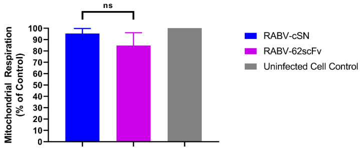
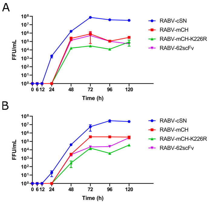
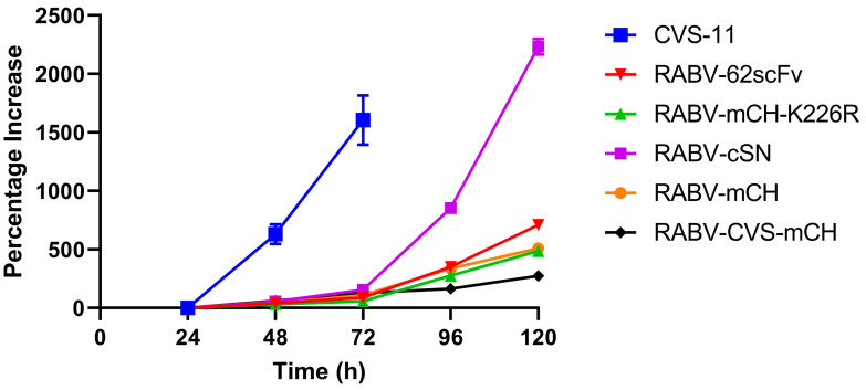
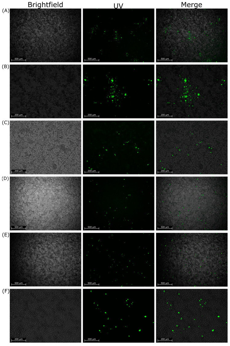
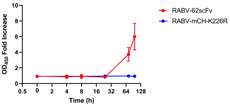
Rabies virus (RABV) causes possibly the oldest disease and is responsible for an estimated >59,000 human fatalities/year. Post exposure prophylaxis (PEP), the administration of vaccine and rabies immunoglobulin, is a highly effective tool which is frequently unavailable in RABV endemic areas. Furthermore, due to the constraints of the blood-brain barrier, current PEP regimes are ineffective after the onset of clinical symptoms which invariably result in death. To circumvent this barrier, a live-attenuated recombinant RABV expressing a highly RABV-neutralising scFv antibody (62-71-3) linked to the fluorescent marker mCherry was designed. Once rescued, the resulting construct (named RABV-62scFv) was grown to high titres, its growth and cellular dissemination kinetics characterised, and the functionality of the recombinant 62-71-3 scFv assessed. Encouraging scFv production and subsequent virus neutralisation results demonstrate the potential for development of a therapeutic live-attenuated virus-based post-infection treatment (PIT) for RABV infection.

Keywords: PEP; antibody; lyssavirus; rabies; rabies treatment; scFv; virus attenuation.


Conflict of interest statement

The authors declare no conflict of interest.

Figures

Figure 1
Figure 1
Assessing the effect of RABV-62scFv infection on mitochondrial respiration. An MTS assay was performed on N2a cells to assess the effect of RABV-62scFv and RABV-cSN infection on N2a cell viability. Output values were compared against the uninfected cell control to obtain a baseline value for mitochondrial respiration. The experiment was undertaken in triplicate to n = 3. Results were analysed using the Brown-Forsythe and Welch ANOVA Test (F* 5.299) where ns p > 0.05.
Figure 2
Figure 2
Growth kinetics of RABV-cSN, RABV-mCH, RABV-mCH-K226R, and RABV-62scFv. Growth kinetics were assessed by multi-step growth curves in both BHK-21 (A) and N2a cells (B). After 1 h infection of either a BHK-21 or N2a cell monolayer at 0.01 MOI, supernatant was taken at 0, 6, 12, 24, 48, 72, 96, and 120 h before FFU/mL was measured by virus titration. Experiment was performed in triplicate to n = 3.
Figure 3
Figure 3
Viral dissemination assay investigating the differences between the cellular dissemination of RABV-cSN, RABV-mCH, RABV-mCH-K226R, RABV-62scFv, CVS-11, and RABV-CVS-mCH over a 120 h period.
Figure 4
Figure 4
Representative images of foci generated in the cellular dissemination assay by either RABV-cSN, RABV-62scFv, or RABV-CVS-mCherry infection. (A,B) show images of foci generated by RABV-cSN infection ((A) at 10×, (B) at 4× magnification). (C,D) show images of foci generated by RABV-62scFv infection at similar magnifications, respectively. (E,F) show images of foci generated by RABV-CVS-mCH infection at similar magnifications, respectively. Images are displayed as brightfield, FITC fluorescence under UV, and a merged image. All cells have been stained for RABV N protein and visualised under UV. All images taken at 96 h post infection.
Figure 5
Figure 5
Charting r62-71-3 scFv production over time after BHK-21 cell culture monolayer infection with RABV-62scFv. Supernatant from RABV-mCH-K226R has been included as a control and results are shown as fold-increase of the negative media-only control. Where possible, error bars representing the standard error of the mean have been shown.
Figure 6
Figure 6
Assessment of the presence of RABV-62scFv to cross a hCMEC/D3 BBB. (A) samples taken from apical and basolateral compartments, respectively, after hCMEC/D3 monolayer infection (72 h) with RABV-62scFv (lanes 2/3) or heat-inactivated RABV-62scFv (lanes 4/5) and amplified for RABV-specific sequences. (B) Kinetics of hCMEC/D3 BBB model formation over time. At each 24 h time point after cell seeding, an apparent permeability (Papp) value was calculated. (C) Images taken of hCMEC/D3 blood-brain barrier monolayer to illustrate cellular infection with RABV-62scFv. Row (i) shows brightfield, UV illumination, and overlay images of hCMEC/D3 cells treated with live RABV-62scFv, while row (ii) shows images taken from hCMEC/D3 cells treated with heat-inactivated RABV-62scFv.
Figure 7
Figure 7
Survival curves of mice used to assess RABV-62scFv toxicity. Mice were given RABV-62scFv, RABV-CVS-mCH, or a media control either ic (A) or iv (B). Mice were then observed for 28 days for the development of clinical symptoms where they would be humanely sacrificed. Dosages of 101, 102, 103, or 104 FFU of virus (or media control) in 30 µL was delivered by needle either into the tail vein (iv) or brain (ic) of 3–4-week-old Balb/c mice.
Figure 8
Figure 8
Serological assessment by mFAVN of mouse sera after challenge (ic or iv) at various FFU concentrations with either RABV-62scFv or RABV-CVS-mCH or media control. (A) shows results from mice challenged ic while (B) shows results from mice challenged iv. Sera from mice given a media control were tested for neutralisation against RABV-CVS-mCH. The dotted line represents the cutoff for detection at a reciprocal antibody titre of 7.49.
Figure 9
Figure 9
Histopathological assessment of mouse brains after ic challenge with RABV-CVS-mCH. Images (AE) show representative images of sections from a mouse brain. Left hand side images have been taken at 200× magnification (AD) or 100× magnification (E), with the right-hand side images showing magnification of the areas bordered by the dotted boxes in the left-hand side images at 400× resolution. (A,B) show the hippocampus region, where brown staining for the RABV N protein is shown in (A) and staining for CD3 is shown in (B). Images (C,D) show midbrain sections where brown staining indicates the presence of RABV N protein and CD3, respectively. Lastly, (E) shows a section of the thalamus where brown staining indicates RABV N staining.

Similar articles

Cited by

References

    1. Nandi S., Kumar M. Development in Immunoprophylaxis against Rabies for Animals and Humans. Avicenna J. Med. Biotechnol. 2010;2:3–21. - PMC - PubMed
    1. Fooks A.R., Cliquet F., Finke S., Freuling C., Hemachudha T., Mani R.S., Muller T., Nadin-Davis S., Picard-Meyer E., Wilde H., et al. Rabies. Nat. Rev. Dis. Primers. 2017;3:17091. doi: 10.1038/nrdp.2017.91. - DOI - PubMed
    1. Hampson K., Coudeville L., Lembo T., Sambo M., Kieffer A., Attlan M., Barrat J., Blanton J.D., Briggs D.J., Cleaveland S., et al. Global Alliance for Rabies Control Partners for Rabies, P. Estimating the global burden of endemic canine rabies. PLoS Negl. Trop Dis. 2015;9:e0003709. - PMC - PubMed
    1. Conzelmann K.K., Cox J.H., Schneider L.G., Thiel H.J. Molecular cloning and complete nucleotide sequence of the attenuated rabies virus SAD B19. Virology. 1990;175:485–499. doi: 10.1016/0042-6822(90)90433-R. - DOI - PubMed
    1. Borca M.V., Ramirez-Medina E., Silva E., Vuono E., Rai A., Pruitt S., Holinka L.G., Velazquez-Salinas L., Zhu J., Gladue D.P. Development of a Highly Effective African Swine Fever Virus Vaccine by Deletion of the I177L Gene Results in Sterile Immunity against the Current Epidemic Eurasia Strain. J. Virol. 2020;94:10–1128. doi: 10.1128/JVI.02017-19. - DOI - PMC - PubMed

Publication types