. 2023 Aug 15;15(8):1744.
doi: 10.3390/v15081744.

Evaluation of Nafamostat as Chemoprophylaxis for SARS-CoV-2 Infection in Hamsters

Affiliations

Evaluation of Nafamostat as Chemoprophylaxis for SARS-CoV-2 Infection in Hamsters

Megan Neary et al. Viruses. .

Abstract

The successful development of a chemoprophylaxis against SARS-CoV-2 could provide a tool for infection prevention that is implementable alongside vaccination programmes. Nafamostat is a serine protease inhibitor that inhibits SARS-CoV-2 entry in vitro, but it has not been characterised for chemoprophylaxis in animal models. Clinically, nafamostat is limited to intravenous delivery and has an extremely short plasma half-life. This study sought to determine whether intranasal dosing of nafamostat at 5 mg/kg twice daily was able to prevent the airborne transmission of SARS-CoV-2 from infected to uninfected Syrian Golden hamsters. SARS-CoV-2 RNA was detectable in the throat swabs of the water-treated control group 4 days after cohabitation with a SARS-CoV-2 inoculated hamster. However, throat swabs from the intranasal nafamostat-treated hamsters remained SARS-CoV-2 RNA negative for the full 4 days of cohabitation. Significantly lower SARS-CoV-2 RNA concentrations were seen in the nasal turbinates of the nafamostat-treated group compared to the control (p = 0.001). A plaque assay quantified a significantly lower concentration of infectious SARS-CoV-2 in the lungs of the nafamostat-treated group compared to the control (p = 0.035). When taken collectively with the pathological changes observed in the lungs and nasal mucosa, these data are strongly supportive of the utility of intranasally delivered nafamostat for the prevention of SARS-CoV-2 infection.

Keywords: SARS-CoV-2; chemoprophylaxis; nafamostat.


Conflict of interest statement

A.O. and S.R. are Directors of Tandem Nano Ltd. M.N., J.H., A.O. and S.R. are co-inventors of patents relating to drug delivery. A.O. has received research funding from ViiV, Merck, Janssen and consultancy from Gilead, ViiV and Merck not related to the current paper. S.R. has received research funding from ViiV and AstraZeneca and consultancy from Gilead not related to the current paper. J.P.S. has received research funding from ENA respiratory, and consultancy from Byotrol Technologies not related to current paper. No other conflicts are declared by the authors.

Figures

Figure 1
Figure 1
Diagrammatic representation of the experimental design employed. Study of nafamostat as chemoprophylaxis against airborne Wuhan SARS-CoV-2 infection. Sentinel hamsters were dosed intranasally twice daily with either 5 mg/kg nafamostat or water for 24 h prior to being cohoused separately in a divided cage with a hamster which was untreated and had been inoculated with Wuhan SARS-CoV-2. All animals were then throat swabbed for 4 days before study termination.
Figure 2
Figure 2
Hamster weights separated by group and inoculation status. All hamsters within each treatment group (n = 15), as well as the donor hamster (inoculated with Wuhan SARS-CoV-2) cohoused with each group (n = 10), were weighed at 24 h intervals up to the study endpoint on day 4. All weights are shown as a percentage of the initial weight recorded at baseline on day −1 of the study. Error bars represent the standard deviation for each study group at each timepoint.
Figure 3
Figure 3
Viral quantification of SARS-CoV-2 N-RNA within lung and nasal turbinate samples obtained from the study groups at day 4. Quantification of SARS-CoV-2 N-RNA in (A) lung and (B) nasal turbinate samples from the Wuhan infected, untreated (donor) hamsters (n = 10), the water-treated hamsters (n = 15) and the nafamostat-treated hamsters (n = 15) at day 4. An unpaired t-test was used to determine statistical significance (p ≤ 0.05) between the water-treated control group and the nafamostat-treated group. ns = not statistically significant. ** = statistically significant (p ≤0.01 and > 0.001).
Figure 4
Figure 4
SARS-CoV-2 RNA quantified from throat swab samples taken from each study group. SARS-CoV-2 viral N-RNA quantified from throat swab samples taken at day 1, 2, 3 and 4 of the study from the (A) Wuhan infected, untreated (donor) control hamsters (n = 10), (B) water-treated hamsters (n = 15) or (C) nafamostat-treated hamsters (n = 15). LOD = limit of detection. A two-way ANOVA was applied to determine statistical significance between the water-treated control group (B) and the nafamostat-treated group (C) at each timepoint; no significant difference was observed (p = 0.5163).
Figure 5
Figure 5
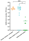
SARS-CoV-2 titres quantified from tissue samples from the donor, nafamostat- or water-treated hamsters. An unpaired t-test was used to determine statistical significance (p ≤ 0.05) between the water-treated control group and the nafamostat-treated group. * = statistically significant (p ≤ 0.05 and >0.001). **** = statistically significant (p ≥ 0.0001).
Figure 6
Figure 6
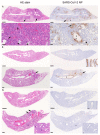
Histological findings and viral antigen expression in lungs and nose of donor; water- or nafamostat-treated hamsters examined at 4 days after intranasal infection of the donor by 104 PFU of SARS-CoV-2 Wuhan. (A) Donor hamster (animal #6), left lung, longitudinal section. The overview of the HE-stained section shows multifocal, bronchiole-centred, consolidated areas (top left: arrows); a higher magnification of the focal area (*) reveals a dense leukocyte infiltration, comprising macrophages, fewer lymphocytes and a variable numbers of neutrophils, with focal desquamation of infected cells (bottom left: arrowheads) and mild hyperplasia of type II pneumocytes/bronchiolar epithelial cells (bottom left: arrows). Immunohistology shows abundant SARS-CoV-2 N expression in patches of alveoli, often in the periphery of consolidated areas (right: arrows). A higher magnification (*) also reveals staining in small patches of unaltered alveoli (bottom right: large arrowhead) and in bronchial epithelial cells (bottom right: small arrowhead). B: Bronchiole. (B) Water-treated sentinel animals. Top: Animal #2 (cage 1; N-RNA and protein negative in lung and nose, plaque assay negative). The lung is histologically unaltered (HE stain, left) and there is no obvious viral antigen expression (right). Inset: Nasal mucosa with focally abundant SARS-CoV-2 N positive epithelial cells. Bottom: Animal #7 (cage 3). The lung exhibits a focal area of peribronchiolar consolidation (left: arrow) with moderate viral antigen expression in bronchus and several bronchioles (right: arrow) as well as in some patches of alveoli. B: Bronchiole. (C) Nafamostat-treated sentinel animals. Top: Animal #6 (cage 7; N-RNA and protein negative in lung and nose, plaque assay negative). The lung is histologically unaltered (HE stain, left), and both lungs (right) and nasal mucosa (inset) do not exhibit viral antigen expression. Bottom: Animal #8 (cage 8; N-RNA and protein negative in lung and nose, plaque assay negative). The lung exhibits a few small, consolidated areas (HE stain, left; arrows); a higher magnification of the focal area (*) reveals hyperplasia of type II pneumocytes/bronchiolar epithelial cells (insets: arrows) with mild leukocyte infiltration. There is no evidence of viral antigen expression (right). B: Bronchiole. (A). Bars = 1 mm (top images) and 50 µm (bottom images). (B). Bars = 1 mm (overview images) and 50 µm (inset, nasal mucosa). (C). Bars = 500 µm (overview images) and 50 µm (insets).

References

    1. Kuzmina A., Khalaila Y., Voloshin O., Keren-Naus A., Boehm-Cohen L., Raviv Y., Avni Y., Rosenberg R., Taube R. SARS-CoV-2 spike variants exhibit differential infectivity and neutralization resistance to convalescent or post-vaccination sera. Cell Host Microb. 2021;29:522–528.e2. doi: 10.1016/j.chom.2021.03.008. - DOI - PMC - PubMed
    1. Yang L., Wang Z. Bench-to-bedside: Innovation of small molecule anti-SARS-CoV-2 drugs in China. Eur. J. Med. Chem. 2023;2023:115503. - PMC - PubMed
    1. Li H., van der Linden W.A., Verdoes M., Florea B.I., McAllister F.E., Govindaswamy K., Elias J.E., Bhanot P., Overkleeft H.S., Bogyo M. Assessing subunit dependency of the Plasmodium proteasome using small molecule inhibitors and active site probes. ACS Chem. Biol. 2014;9:1869–1876. doi: 10.1021/cb5001263. - DOI - PMC - PubMed
    1. Godbole A.A., Ahmed W., Bhat R.S., Bradley E.K., Ekins S., Nagaraja V. Targeting Mycobacterium tuberculosis topoisomerase I by small-molecule inhibitors. Antimicrob. Agents Chemother. 2015;59:1549–1557. doi: 10.1128/AAC.04516-14. - DOI - PMC - PubMed
    1. Christ F., Shaw S., Demeulemeester J., Desimmie B.A., Marchand A., Butler S., Smets W., Chaltin P., Westby M., Debyser Z., et al. Small-molecule inhibitors of the LEDGF/p75 binding site of integrase block HIV replication and modulate integrase multimerization. Antimicrob. Agents Chemother. 2012;56:4365–4374. doi: 10.1128/AAC.00717-12. - DOI - PMC - PubMed

Publication types