Review
. 2023 Oct;10(30):e2303259.
doi: 10.1002/advs.202303259. Epub 2023 Aug 26.

Nitric Oxide: Physiological Functions, Delivery, and Biomedical Applications

Affiliations
Review

Nitric Oxide: Physiological Functions, Delivery, and Biomedical Applications

Syed Muntazir Andrabi et al. Adv Sci (Weinh). 2023 Oct.

Abstract

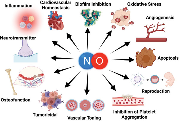
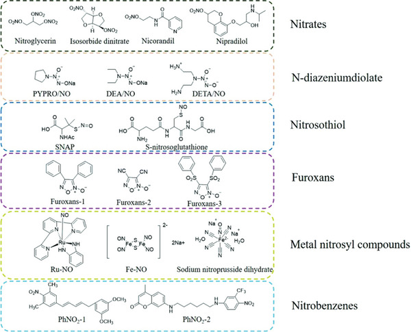
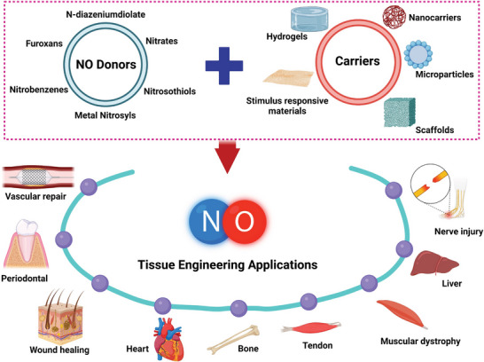
Nitric oxide (NO) is a gaseous molecule that has a central role in signaling pathways involved in numerous physiological processes (e.g., vasodilation, neurotransmission, inflammation, apoptosis, and tumor growth). Due to its gaseous form, NO has a short half-life, and its physiology role is concentration dependent, often restricting its function to a target site. Providing NO from an external source is beneficial in promoting cellular functions and treatment of different pathological conditions. Hence, the multifaceted role of NO in physiology and pathology has garnered massive interest in developing strategies to deliver exogenous NO for the treatment of various regenerative and biomedical complexities. NO-releasing platforms or donors capable of delivering NO in a controlled and sustained manner to target tissues or organs have advanced in the past few decades. This review article discusses in detail the generation of NO via the enzymatic functions of NO synthase as well as from NO donors and the multiple biological and pathological processes that NO modulates. The methods for incorporating of NO donors into diverse biomaterials including physical, chemical, or supramolecular techniques are summarized. Then, these NO-releasing platforms are highlighted in terms of advancing treatment strategies for various medical problems.

Keywords: biomedical applications; delivery; donors; nitric oxide; physiological functions.


Conflict of interest statement

The authors declare no conflict of interest.

Figures

Figure 1
Figure 1
Major physiological roles mediated by exogenously and endogenously produced NO. Figure created using BioRender.com.
Figure 2
Figure 2
Schematic representation showing the structure of the several classes of NO donors categorized into specific groups. Reproduced with permission.[ 173b ] Copyright 2021, under Creative Commons Attribution License (CC BY).
Figure 3
Figure 3
The role of NO in cell migration and wound closure. A) MIP‐177 and NO loaded MIP‐177 effect on wound closure in in‐vitro for 48 h. B) Microscopic images showing the pre‐migration stage without detection mask at time = 0 h and the migration of cells detected with a mask after 48 h. The black circles indicate the presence of MIP‐177 particles between the migrated cells. Reproduced with permission.[ 207b ] Copyright 2020, John Wiley and Sons Inc.
Figure 4
Figure 4
Effect of NO‐releasing scaffold on angiogenesis and wound healing. A) Representative images showing neovascularization and respective CD31 expression (blood vessels stained in green). B) Presents quantitative data of the length of vessels and C) CD31 stained cells, respectively. D) Digital images showing the effect on wound healing by different groups and E) Corresponds to wound remaining area at preset time points. F–J) Gene expression of in vivo wound tissue samples on days 7 and 13 by western blotting analysis. Adapted with permission.[ 208 ] Copyright 2020, American Chemical Society. Quantitative analysis of growth factors in new skin tissues of wound K) NO, a repair factor, L) Hydroxyproline, for collagen deposition, M) CD31, N) TGF‐β, and O) VEGF. Reproduced with permission.[ 212 ] Copyright 2022, Elsevier Ltd.
Figure 5
Figure 5
Photothermal effect and NO‐based antibacterial activity. A) Loading ratio (%) of BNN6 (NO donor) in GO‐based vehicles. B) NIR‐based color change of GO‐βCD in the absence and presence of BNN6. C) Release of NO with or without NIR irradiations. D‐G) Agar plates show the antibacterial activity of GO‐βCD‐BNN6 and other groups along with their respective quantitative bacterial viability against S. aureus (D,E) and E. coli (F,G), respectively. Reproduced with permission.[ 226 ] Copyright 2020, American Chemical Society.
Figure 6
Figure 6
In‐vivo antibacterial and wound healing efficacy of DMAH. A) Antibacterial activity against MRSA on day 2. B) Digital images display wound healing at desired time points after treatment with respective groups and C) Presents the quantification of wound closure on days 2, 5, 10, and 15. D) H&E staining of skin tissue from the wound showing wound healing and neutrophil infiltration (green circles and arrows). All Scale bars are 20 µm. Adapted with permission.[ 227 ] Copyright 2022, John Wiley and Sons Inc.
Figure 7
Figure 7
Concentration‐dependent antimicrobial activity of NO under laser irradiation. A,B) Bacterial viability of E. coli and S. aureus respectively, treated with Fe3O4@PDA@PAMAM‐G3 and Fe3O4@PDA@PAMAM@NONOate under different laser irradiation conditions. C) Bacterial colonies formation of E. coli and S. aureus under different treatments. D) Bacterial colonies formation of E. coli and S. aureus treated with different concentrations of Fe3O4@PDA@PAMAM@NONOate. Reproduced with permission.[ 228b ] Copyright 2018, John Wiley and Sons Inc.
Figure 8
Figure 8
Effect of NO‐releasing scaffolds on platelet adhesion and activation. A) Activated platelets in red (CD62p stain) and corresponding SEM images in PCL/PCL‐MOFs‐GSNO. B) Quantitative estimation of platelet adhesion. C) Digital image presenting ex‐vivo AV shunt model. D,E) Thrombus formation (mg) and adhered platelet count in PCL/PCL‐MOFs‐GSNO scaffolds, respectively. Scale bar: 20 µm. Reproduced with permission.[ 234 ] Copyright 2021, Elsevier Inc.
Figure 9
Figure 9
NO containing stent‐cell interaction and in‐vivo implantation. A,B) Digital images of the vascular stent in cross‐sectional view after exposure to blood flow for 2 h and thrombus formation on corresponding foils, respectively. C,D) Effect of developed HA@DOTA‐Cu and other vascular grafts on smooth muscle cell migration and HUVECs, respectively. E) Fluorescent images show cell integration on vascular stent of 316L SS (control) and HA@DOTA‐Cu after implantation. F) Histomorphometric analysis of implanted stents in‐vivo. Reproduced under Creative Commons Attribution License (CC BY).[ 241 ] Copyright 2021, The Authors. Published by Wiley‐VCH.
Figure 10
Figure 10
Schematic illustration of the enhanced tumor therapy with reversed multidrug resistance (MDR) tumor cells and inhibited metastasis by drug co‐delivery and in situ vascular‐promoting strategy. The compounds under test are d‐α‐tocopherol polyethylene 1000 glycol succinate (TPGS), TPGS‐paclitaxel (TPGS‐SS‐PTX or TSP), TPGS derived NO donor (TPGS‐NO3 or TN), and micellar TPGS‐SS‐PTX and TPGS‐NO3 (TSP‐TN). TSP‐TN not only caused vasodilation and angiogenesis reducing leaky vasculature but also induced apoptosis in MDR tumor cells. Reproduced with permission.[ 244b ] Copyright 2017, Elsevier Inc.
Figure 11
Figure 11
In vivo antitumor efficacy against MDR tumors. A) Change in tumor (MCF‐7/ADR) volume profile, B) Tumor weight, C) Relative body weight, and D) Tunnel assay to evaluate apoptosis of tumor cells in mice *p < 0.05, **p < 0.01, and ***p < 0.001 versus TSP. E) Blood vessels (α‐CD31 antibody stained as red and DAPI (blue) for nuclei) and tumor apoptosis (TUNEL staining as green) presented by immune‐fluorescent images. F) Digital images of dissected lungs in B16F10 metastatic model. G,H) Number and diameter of nodules in metastatic lungs. *p < 0.05, **p < 0.01, and ***p < 0.001 versus saline. #p < 0.05, ##p < 0.01, and ###p < 0.001 versus Taxol. I) H&E images of metastatic lung sections. Reproduced with permission.[ 244b ] Copyright 2017, Elsevier Inc.
Figure 12
Figure 12
NO delivery for tissue repair and regeneration.
Figure 13
Figure 13
NO promotes the regeneration of nerves, muscle, and bone. A–C) Quantitative data showing enhanced revascularization, motor functioning, and nerve regeneration respectively, with NO‐SN in nerve crush injury rat model. Reproduced with permission under Creative Commons Attribution License (CC BY).[ 256 ] Copyright 2021, The Authors. Published by Wolters Kluwer. D) H&E staining of muscle sections from the gastrocnemius injury site showed inflammatory and more fibrotic tissue formation in L‐NAME treated group (iNOS inhibitor) after 24 h compared to sham and trauma groups. E,F) Increased collagen deposition in L‐NAME treated group after 7 days as compared to other groups showed by Sirius red staining alone (E) and under polarized light (F). Reproduced with permission.[ 259a ] Copyright 2010, Elsevier Inc. Panels (G,H) show the scheme of Osteoporosis treatment using NIR‐regulated UCPA‐BNN gas therapy in the OVX model. Panel (I) shows the microarchitecture of trabecular bone using micro‐CT for the third lumbar vertebral body and the distal femur of each group. Panels (J) and (K) represent the quantitative data of architectural parameters of BMD (bone mineral density) and Tb. N (trabecular number) respectively, in OVX mice. Reprinted with permission.[ 266 ] Copyright 2021, American Chemical Society.

References

    1. a) Snyder S. H., Bredt D. S., Sci. Am. 1992, 266, 68; - PubMed
    2. b) Bredt D. S., Free Radic. Res. 1999, 31, 577. - PubMed
    1. a) Ignarro L. J., J Card Surg 2002, 17, 301; - PubMed
    2. b) Palmer R. M., Ferrige A. G., Moncada S., Nature 1987, 327, 524. - PubMed
    1. O'Dell T. J., Hawkins R. D., Kandel E. R., Arancio O., Proc. Natl. Acad. Sci. USA 1991, 88, 11285. - PMC - PubMed
    1. Miller M. R., Megson I. L., Br. J. Pharmacol. 2007, 151, 305. - PMC - PubMed
    1. Gow A. J., Farkouh C. R., Munson D. A., Posencheg M. A., Ischiropoulos H., Am. J. Physiol. Lung Cell Mol. Physiol. 2004, 287, L262. - PubMed

Publication types

LinkOut - more resources