. 2024 Jun 3;147(6):2098-2113.
doi: 10.1093/brain/awad280.

Toxic effects of mutant huntingtin in axons are mediated by its proline-rich domain

Affiliations

Toxic effects of mutant huntingtin in axons are mediated by its proline-rich domain

Scott T Brady et al. Brain. .

Erratum in

Abstract

Huntington's disease results from expansion of a polyglutamine tract (polyQ) in mutant huntingtin (mHTT) protein, but mechanisms underlying polyQ expansion-mediated toxic gain-of-mHTT function remain elusive. Here, deletion and antibody-based experiments revealed that a proline-rich domain (PRD) adjacent to the polyQ tract is necessary for mHTT to inhibit fast axonal transport and promote axonal pathology in cultured mammalian neurons. Further, polypeptides corresponding to subregions of the PRD sufficed to elicit the toxic effect on fast axonal transport, which was mediated by c-Jun N-terminal kinases (JNKs) and involved PRD binding to one or more SH3-domain containing proteins. Collectively, these data suggested a mechanism whereby polyQ tract expansion in mHTT promotes aberrant PRD exposure and interactions of this domain with SH3 domain-containing proteins including some involved in activation of JNKs. In support, biochemical and immunohistochemical experiments linked aberrant PRD exposure to increased JNK activation in striatal tissues of the zQ175 mouse model and from post-mortem Huntington's disease patients. Together, these findings support a critical role of PRD on mHTT toxicity, suggesting a novel framework for the potential development of therapies aimed to halt or reduce axonal pathology in Huntington's disease.

Keywords: Huntington’s disease; JNK3; SH3-binding domain; axonal transport; huntingtin; proline-rich domain.


Conflict of interest statement

The authors report no competing interests

Figures

Figure 1
Figure 1
Toxicity of huntingtin fragments on fast axonal transport. (A) Huntingtin (HTT) is a protein of unknown function containing more than 3000 amino acids. The absence of an identified catalytic activity suggests a role for HTT as a scaffold for unknown cellular components. While previous studies have determined that exon 1 is sufficient for neurotoxicity, HTT contains multiple potential protein interaction domains [e.g. polyQ tract, a proline-rich domain (PRD) and HEAT repeats], which could provide additional biological activities. Three different recombinant mHTT fragments ranging from approximately 100 to 1000 amino acids were generated to determine whether different mHTT fragments exhibit different toxicities on fast axonal transport in the isolated axoplasm model. (B) All three mutant HTT (mHTT) constructs perfused at 100 nM have the same inhibitory effect on both anterograde and retrograde fast axonal transport rates. #P < 0.0001; *P < 0.001. (C) Exon 1 of mHTT appears to contain the toxic element causing the inhibitory effect in axons. There are two major motifs in exon 1: the expanded polyQ tract and a PRD. Previous studies generated monoclonal antibodies (Abs) to polyQ (MW1) and to polyP (MW7). (D and E) Plots depicting results from vesicle motility assays in isolated squid axoplasm. mHTT exon 1 (49Q) was perfused into axoplasm (100 nM) and fast axonal transport rates monitored by video microscopy, as in B. Individual rate measurements (µm/s, arrowheads) are plotted as a function of time (min). Both anterograde (black arrowheads and lines) and retrograde (reverse grey arrowheads and lines) fast axonal transport rates are shown. (D) Preincubation of mHTT exon 1 (49Q) with MW1 Ab (polyQ-directed) did not alter the toxic effects of this protein, so both anterograde and retrograde transport were still inhibited. n = number of axoplasms evaluated. (E) In contrast, preincubation with MW7 (polyP directed) completely blocked the effects of HTT exon 1 (Q49) on axonal transport. This suggests that exposure and/or accessibility of the PRD is essential for mHTT toxicity on fast axonal transport. (F) Box plots show that preincubation with M1 Ab did not significantly alter the toxic effects of mHTT exon 1 (Q49) on either anterograde or retrograde fast axonal transport. In contrast, preincubation with MW7 significantly reduced such effects. #P < 0.0001.
Figure 2
Figure 2
Deletion of the PRD eliminate toxic effects of mHTT fragments on fast axonal transport. (A) Perfusion of mHTT exon 1 (Q49) at 100 nM inhibits fast axonal transport in both directions through activation of the MAPK JNK3, (B) The same mHTT construct with the proline-rich domain (PRD) deleted [mHTT exon 1 (Q49 ΔPRD)] has no effect on axonal transport in either direction at 100 nM. (C and D) Similar effects are seen with a longer mHTT (1–969 Q46) fragment at 100 nM. Deletion of the PRD [mHTT (1–969 Q46 ΔPRD)] eliminates the toxic effects on fast axonal transport elicited by mHTT (1–969 Q46). (E) Quantitative comparison of rates observed between 30 and 50 min with each construct shows that the PRD is necessary for the toxicity of both mHTT exon 1 (Q49) and mHTT (1–969 Q46). #P < 0.0001. (F) A glutathione-S-transferase (GST) tagged protein in which the PRD from HTT is added to the C-terminus (GST-PRD) has the same effect on both anterograde and retrograde axonal transport without the presence of a polyQ stretch. The inhibitory effect of GST-PRD is not significantly different from the effects of pathogenic forms of HTT with an expanded polyQ. This suggests that the PRD is both necessary and sufficient to produce the effect of pathological mutant HTT on fast axonal transport. n = number of axoplasms.
Figure 3
Figure 3
Toxic effects of mHTT-exon 1 (Q46) on axons of primary cultured cortical neurons depend on the PRD. (A) Cortical neurons, transfected to express mCherry (red), were also visualized with fluorescent membrane-permeant marker NeuO, a live neuron-specific dye (green), and a DNA label (Hoechst, in blue). (A) Cortical neurons were transfected with mCherry empty expression vector (mCherry, control); (B) mCherry fused to mHTT exon 1 (Q46) [mCherry-mHTT exon 1 (Q46)]; or (C) mCherry fused to mHTT exon 1 (Q46) with proline-rich domain (PRD) deletion [mCherry-mHTT exon 1 (Q46 ΔPRD)]. Cortical neurons expressing mCherry-mHTT exon 1 (Q46) displayed membrane/cytoplasmic morphological disturbances in both neurites (B, arrow) and soma (B, arrowheads), whereas neurons expressing mCherry-mHTT exon 1 (Q46 ΔPRD) did not. Scale bars = 50 μm. See also Supplementary Fig. 1.
Figure 4
Figure 4
Reduced outgrowth and degeneration of axons from primary cortical neurons following transfection with cherry-tagged mHTT exon 1, but not with cherry-tagged mHTT exon 1 lacking the proline-rich domain (PRD). Live imaging of axons elongating within the microchannels of our custom microfluidic chambers were visualized with the live neuronal marker, NeuO, beginning just prior to transfection at 6 days in vitro (DIV) after many axons have already entered the microchannels (Supplementary Fig. 2) and continuing until 13 DIV, or 7 days post-transfection. This design evaluates the effect of constructs on the growth and viability of pre-existing axons. Neurons were transfected with (A) mCherry-mHTT exon 1 (Q46) or with (B) mCherry-mHTT exon 1 (Q46 ΔPRD). The length of axons within each microchannel was measured as they entered the microchannel from the somatodendritic compartment (SDC) until it reached the axon terminal compartment (ATC). Cortical neurons expressing mCherry-mHTT exon 1 (Q46) developed axonal discontinuities, observed with the loss of NeuO labelling (A, arrows), illustrating axonal degeneration within the microchannel and in the ATC. There was an apparent reduction of cortical neuron somas in the SDC for cultured neurons transfected with mCherry-mHTT exon 1 (Q46) (A, arrowhead), but degeneration of neuronal soma was not quantitatively evaluated. Scale bar = 50 μm. (C) The total number of axons elongating and degenerating in cortical neurons expressing mHTT exon 1 (Q46) (n = 433) or mHTT exon 1 (Q46 ΔPRD) (n = 412) was quantified over time. Scale bars = 50 μm. Analysis of 261 axons of untransfected neurons had no degenerating axons. See also Supplementary Figs 2 and 3. (D) Comparison of axonal degeneration between neurons expressing mCherry-mHTT exon 1 (Q46) and mCherry-mHTT exon 1 (Q46 ΔPRD) indicates that the fraction of degenerating axons was reduced by >90% (P = 0.0067).
Figure 5
Figure 5
Aberrant activation of JNKs and increased PRD exposure in the zQ175 mouse model of Huntington’s disease. (A) Immunoblots of striatal protein lysates prepared from wild type (n = 4) and heterozygous zQ175 (n = 5) mice were analysed with antibodies that recognize kinesin heavy chains (KHC), the striatum-enriched protein DARPP-32, total (total JNK) and active (pJNK) forms of JNKs, and synaptophysin (synaptoph). (B) Li-COR quantitation of immunoreactive bands in A revealed that, compared to wild-type mice, zQ175 mice striata has lower levels of striatal DARPP32 (DARPP32/KHC ratios), increased activation of JNKs (total JNK/pJNK ratios), and similar levels of synaptophysin (synaptoph/KHC ratios). (CE) Immunohistochemical analyses of striatal tissues from YFP (control) and YFP-zQ175 mice littermates (n = 4 mice/genotype). Nuclei were stained using DAPI (in blue) in all panels. (C) An antibody that recognizes an epitope close to the P1218 residue in HTT (D7F7, in red) displayed increased immunoreactivity in YFP mice, compared to YFP-zQ175 mice littermates. This finding was confirmed by quantitative dot blot experiments in Supplementary Fig. 4A, suggesting reduced levels of total HTT expression in zQ175 mice, compared to wild-type (WT) mice. (D and E) Conversely, and despite this reduction, MW7 antibody immunoreactivity was markedly increased in striata of YFP-zQ175 mice, compared to YFP mice littermates. Results from quantitative dot blot experiments in Supplementary Fig. 4C supported and extended this finding. Collectively, data in CE suggested increased exposure of the PRD in mHTT, compared to wild-type HTT.
Figure 6
Figure 6
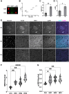
Aberrant activation of JNKs and increased PRD exposure in striatal tissue from HD patients. (A) A representative dot blot of human postmortem caudate tissue (see Supplementary Table 2 for patient demographic information) showed increased immunoreactivity of MW7 and pJNK in Huntington’s disease (HD) patients, despite a moderate reduction in the total level of HTT protein as detected by D7F7 antibody. (B) Quantification of dot blots was plotted for each case (n = 3 for control and n = 3 for HD) to demonstrate a correlation between MW7/D7F7 ratio and JNK activity in HD cases, with controls clustered at the lower quadruple. (C and D) Relative immunoreactivity of MW7 (shown as ratio of MW7/D7F7) and pJNK from the same blots was quantified and plotted to show increases in HD as compared with the control samples (**P = 0.0068, *P = 0.0276, unpaired two-sample T-test). (E) Representative confocal images of the striatum indicated associated and increased immunostaining of MW7 and pJNK in HD patients compared with their controls. Tissues were counterstained with Hoechst as a nuclear marker and Tubb3 as a neuronal marker. (F and G) Quantification was performed on whole striatum images stitched from single images captured by wide-field epifluorescence imaging and showed higher MW7 and pJNK immunoreactivities in HD patients (n = 11) than in controls (n = 5) and the differences were statistically significant (****P < 0.0001, **P < 0.01, *P < 0.05 Kruskal-Wallis one-way ANOVA). HD cases were graded according to neuropathology (see Supplementary Table 3 for patient demographic information of cases examined in EG).
Figure 7
Figure 7
Subdomains P1 and P3 of the PRD suffice to inhibit fast axonal transport via JNKs. (A) The proline-rich domain (PRD) contains three subregions previously shown to interact with SH3 domain- and WW motif-containing proteins: a variable length polyP (P1), a proline rich sequence that also contains Q, L and A residues (P2) and a second polyP stretch (P3). GST-tagged versions of these subregions were perfused in axoplasm and their effect on fast axonal transport evaluated using vesicle motility assays as in Fig. 1. (B) Perfusion of a GST-P1 (100 nM) construct inhibited both anterograde and retrograde fast axonal transport rates. (C) In contrast, a GST-P2 (100 nM) construct has no effect on either direction of fast axonal transport. (D) Effects of the GST-P3 (100 nM) construct on fast axonal transport are comparable to P1, the full-length PRD alone (Fig. 1F) and HTT exon 1 Q49 with an intact PRD (Fig. 1). (E) SP600125 (500 nM), a highly specific pharmacological inhibitor of JNKs, blocked the effect of GST-P3 (100 nM) on transport, just as it does for mHTT exon 1 (Q49) or other pathogenic mHTT fragments. (F) Quantitative comparisons for effects of PRD subdomains on axonal transport. Effects of each peptide on transport were compared to effects of the control wild-type HTT 1–548 (Q23) polypeptide. GST-P1 and GST-P3 significantly inhibited both anterograde and retrograde fast axonal transport as shown in the box plots (P < 0.0001) and the slopes in B and D reflecting a decline in transport. In contrast, GST-P2 did not differ significantly from mHTT (548) Q23 in anterograde transport (P = 0.2599). Although retrograde fast axonal transport was lower than with HTT 1–548 (Q23) (P = 0.0014) (see Figure 1B), there was no decline in retrograde transport rate and GST-P2 started out less that HTT 1–548 (Q23) retrograde, so effects of GST-P2 on axonal transport are likely negligible. Inhibition of JNK MAPK activity by SP600125 prevented the effect so GST-P3 on fast axonal transport. #P < 0.0001, *P < 0.001.
Figure 8
Figure 8
Toxic effects of mHTT on fast axonal transport involve binding to SH3 domain-containing proteins. Proline-rich domains are implicated in a variety of protein–protein interaction domains. (A) SH3-binding domains are a common motif with a characteristic PXXP conserved sequence. The PRD contains multiple PXXP motifs, so we co-perfused mHTT exon 1 (Q49) (100 nM) with a peptide containing an SH3 motif at 1 µM. Excess SH3 peptide prevents mHTT exon 1 (Q49) effects on fast axonal transport. (B) In contrast, a mutant SH3 domain that no longer binds PXXP motifs (SH3-Y52A) at 1 µM fails to protect fast axonal transport. (C) Preincubation with a recombinant peptide encoding an SH3 motif blocked effects of mHTT exon 1 (Q49) on both anterograde and retrograde transport rates (P < 0.0001), whereas a mutant SH3 peptide unable to bind SH3-binding motifs (Y52A) did not (P = 0.41 for anterograde rates; P = 0.1964 for retrograde rates). #P < 0.0001.

Comment in

References

    1. The Huntington's Disease Collaborative Research Group . A novel gene containing a trinucleotide repeat that is expanded and unstable on Huntington’s disease chromosomes. Cell. 1993; 72:971–983. - PubMed
    1. Gusella JF, MacDonald ME. Molecular genetics: Unmasking polyglutamine triggers in neurodegenerative disease. Nat Rev Neurosci. 2000;1:109–115. - PubMed
    1. Gatto RG, Chu Y, Ye AQ, et al. Analysis of YFP(J16)-R6/2 reporter mice and postmortem brains reveals early pathology and increased vulnerability of callosal axons in Huntington’s disease. Hum Mol Genet. 2015;2:5285–5298. - PMC - PubMed
    1. Han I, You Y, Kordower JH, Brady ST, Morfini GA. Differential vulnerability of neurons in Huntington’s disease: The role of cell type-specific features. J Neurochem. 2010;113:1073–1091. - PMC - PubMed
    1. Pouladi MA, Morton AJ, Hayden MR. Choosing an animal model for the study of Huntington’s disease. Nat Rev Neurosci. 2013;14:708–721. - PubMed

MeSH terms