. 2024 Jan 27;45(4):255-264.
doi: 10.1093/eurheartj/ehad562.

Critical limb-threatening ischaemia and microvascular transformation: clinical implications

Affiliations

Critical limb-threatening ischaemia and microvascular transformation: clinical implications

Santeri Tarvainen et al. Eur Heart J. .

Abstract

Background and aims: Clinical management of critical limb-threatening ischaemia (CLTI) is focused on prevention and treatment of atherosclerotic arterial occlusions. The role of microvascular pathology in disease progression is still largely unspecified and more importantly not utilized for treatment. The aim of this explorative study was to characterize the role of the microvasculature in CLTI pathology.

Methods: Clinical high-resolution imaging of CLTI patients (n = 50) and muscle samples from amputated CLTI limbs (n = 40) were used to describe microvascular pathology of CLTI at the level of resting muscle blood flow and microvascular structure, respectively. Furthermore, a chronic, low arterial driving pressure-simulating ischaemia model in rabbits (n = 24) was used together with adenoviral vascular endothelial growth factor A gene transfers to study the effect of microvascular alterations on muscle outcome.

Results: Resting microvascular blood flow was not depleted but displayed decreased capillary transit time (P < .01) in CLTI muscles. Critical limb-threatening ischaemia muscle microvasculature also exhibited capillary enlargement (P < .001) and further arterialization along worsening of myofibre atrophy and detaching of capillaries from myofibres. Furthermore, CLTI-like capillary transformation was shown to worsen calf muscle force production (P < .05) and tissue outcome (P < .01) under chronic ischaemia in rabbits and in healthy, normal rabbit muscle.

Conclusions: These findings depict a progressive, hypoxia-driven transformation of the microvasculature in CLTI muscles, which pathologically alters blood flow dynamics and aggravates tissue damage under low arterial driving pressure. Hypoxia-driven capillary enlargement can be highly important for CLTI outcomes and should therefore be considered in further development of diagnostics and treatment of CLTI.

Keywords: Angiogenesis; Capillaries; Chronic limb-threatening ischaemia; Growth factor; Hypoxia; Ischaemia; Microvascular remodelling; Peripheral arterial disease; Vascular endothelial.


Figures

Structured Graphical Abstract
Hypoxic capillary enlargement is a major problem in chronic limb-threatening ischaemia (CLTI).
Figure 1
Figure 1
Study design and patient characteristics. The study is composed of three parts involving: (i) imaging of resting limb muscles in critical limb-threatening ischaemia (CLTI) patients scheduled for urgent revascularization and healthy volunteers to initially establish the paradoxical conservation of tissue blood flow under critical ischaemia, (ii) muscle sample collection from amputated CLTI limbs to characterize the structural alterations of muscle capillaries under different levels of ischaemic exposure, (iii) modelling the effects of capillary enlargement and arterialization under low arterial driving pressure in a rabbit model of chronic limb ischaemia. The characteristics of CLTI patients recruited to the study are presented in the table. CEU, contrast-enhanced ultrasound; PAI, photoacoustic imaging; PET, [15O]-H2O positron emission tomography; Histo, immunohistology; qPCR, quantitative polymerase chain reaction; Denudation, ischaemia induction via femoral artery denudation; GT, gene transfer; Force, measurement of calf muscle force production; ABI, ankle brachial index; CLTI, critical limb-threatening ischaemia
Figure 2
Figure 2
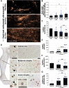
Altered microvascular blood flow kinetics and progressive capillary arterialization in critical limb-threatening ischaemia (CLTI) muscle. (A) Intravascularly delivered contrast signal (spekles) detected by ultrasound imaging was used to assess resting muscle blood flow in CLTI patients and volunteers. (B) Quantitation of contrast-enhanced ultrasound (CEU) signal intensity showed no significant difference in resting muscle blood flow between CLTI patients or volunteers but rather a trend towards increased signal in the ischaemic legs as compared to healthy controls. (C) Capillary transit time of the CEU contrast agent was significantly decreased in ischaemic muscle as compared to volunteers. (D) Structural transformation of muscle capillaries (arrowheads) along worsening of myofibre atrophy was seen in CD31 (brown) immunostanings. Double staining for CD31 + α-SMA (blue and brown, respectively) was used to confirm the presence of α-SMA positive pericytes on arterialized capillaries. (E) Increased mean capillary area, (F) decreased capillary density, and (G) increased capillary size distribution were detected along worsening of myofibre atrophy. Isc, ischaemic leg; Pre, before revascularization; Ctrl, contralateral leg of a patient; Post, after revascularization; CLTI, critical limb-threatening ischaemia; CEU, contrast-enhanced ultrasound. Scale bars 50μm. *P < .05, **P < .01, ***P < .001
Figure 3
Figure 3
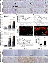
VEGF-A immunostaining has altered pattern in ischaemic muscle and associates with capillary enlargement. Quantification of average mRNA expressions via qPCR from amputation muscle samples displayed increased (A) HIF1A and (B) EPAS1 levels consistent with increased hypoxia but decreased (C) VEGFA expression in the ischaemic samples. (D) Amputation muscles samples from control areas showed distinct patches of VEGF-A −/+ (brown) myofibres. (E) Myofibres in the ischaemic amputation samples displayed heterogeneous VEGF-A expression (brown). (F) CD31 (blue) + VEGF-A (brown) double immunostainings display enlarged capillaries near myofibres that strongly expressed VEGF-A. Scale bars 50 μm. *P < .05
Figure 4
Figure 4
Capillary enlargement by AdVEGF-A under low arterial driving pressure aggravates ischaemic tissue damage. (A) Structural transformation of muscle capillaries (arrowheads) along worsening of myofibre atrophy seen in CD31 (brown) immunostanings 4 weeks in AdLacZ control transduced muscles and further capillary enlargement and arterialization induced by AdVEGF-A. Increased (B) mean capillary area and (C) capillary size distribution were quantified along worsening of myofibre atrophy. (D) Mean contrast-enhanced ultrasound (CEU) signal intensity did not significantly differ between gene transfer groups or as compared to normal (d0) values. (E) Mean CEU arrival time was significantly increased after the ischaemia operation but did not differ between the gene transfer groups. (F) Contrast-enhanced ultrasound signal (spekles) distribution in the transduced muscles (dotted line) was uneven after AdVEGF-A gene transfer leaving parts of the muscle unperfused (asterisks). (G) Calf muscle force production was reduced by the ischaemia operation at the time of gene transfer and further decreased after AdVEGF-A gene transfer. (H) Area of atrophic muscle damage increased after AdVEGF-A gene transfer. (I) Rounding, occasional atrophy, and increased expression of Hif-1a and Hif-2a of myofibres 1 week after AdVEGF-A gene transfer to healthy rabbit skeletal muscle as compared to changes in respective AdLacZ control. CEU, contrast-enhanced ultrasound; d0, baseline; GT, gene transfer; GT + X, X weeks after GT. Scale bar in main images 50μm, inset 25μm. *P < .05, **P < .01, ***P < .001
Figure 5
Figure 5
Microvascular blood flow dynamics as well as oxygen diffusion may be compromised in critical limb-threatening ischaemia (CLTI). (A) Decreased arterial driving pressure can predispose tissues with capillary size heterogeneity to shunting. Where capillaries with uniform diameter and resistance are logically equally perfused, capillary size heterogeneity alters the amount of flow in each capillary. Upon decreased arterial driving pressure, capillary size heterogeneity-mediated shunting may leave normal, high-resistance capillaries unperfused. (B) Capillaries normally are in thigh contact with myocytes. Capillaries in atrophic CLTI muscle, however, are often seen having lost contact with their surrounding myofibres potentially influencing oxygen diffusion. (C) Whilst in normal capillaries the contact surface of erythrocytes and capillary endothelial cells is maximized by squeezing of erythrocytes through the capillary tube, the enlarged capillaries may allow relatively free flow of erythrocytes decreasing contact surface. A decrease in cell membrane contacts could impair oxygen diffusion as diffusion through cell membrane phospholipids has been shown more efficient than diffusion through biological fluids. CLTI, critical limb-threatening ischaemia

Comment in

References

    1. Aboyans V, Ricco J-B, Bartelink M-LEL, Björck M, Brodmann M, Cohnert T, et al. . 2017 ESC Guidelines on the diagnosis and treatment of peripheral arterial diseases, in collaboration with the European Society for Vascular Surgery (ESVS). Eur Heart J 2018;39:763–816. 10.1093/eurheartj/ehx095 - DOI - PubMed
    1. Criqui MH, Matsushita K, Aboyans V, Hess CN, Hicks CW, Kwan TW, et al. . Lower extremity peripheral artery disease: contemporary epidemiology, management gaps, and future directions: a scientific statement from the American Heart Association. Circulation 2021;144:E171–91. 10.1161/CIR.0000000000001005 - DOI - PMC - PubMed
    1. Duff S, Mafilios MS, Bhounsule P, Hasegawa JT. The burden of critical limb ischemia: a review of recent literature. Vasc Health Risk Manag 2019;15:187–208. 10.2147/VHRM.S209241 - DOI - PMC - PubMed
    1. Lindner JR, Womack L, Barrett EJ, Weltman J, Price W, Harthun NL, et al. . Limb stress-rest perfusion imaging with contrast ultrasound for the assessment of peripheral arterial disease severity. JACC Cardiovasc Imaging 2008;1:343–50. 10.1016/j.jcmg.2008.04.001 - DOI - PMC - PubMed
    1. Burchert W, Schellong S, van den Hoff J, Meyer GJ, Alexander K, Hundeshagen H. Oxygen-15-water PET assessment of muscular blood flow in peripheral vascular disease. J Nucl Med 1997;38:93–8. - PubMed

Substances