. 2023 Oct:587:109871.
doi: 10.1016/j.virol.2023.109871. Epub 2023 Aug 19.

Creation and characterization of a recombinant mammalian orthoreovirus expressing σ1 fusion proteins encoding human epidermal growth factor receptor 2 peptides

Affiliations

Creation and characterization of a recombinant mammalian orthoreovirus expressing σ1 fusion proteins encoding human epidermal growth factor receptor 2 peptides

Nicole A Jandick et al. Virology. 2023 Oct.

Abstract

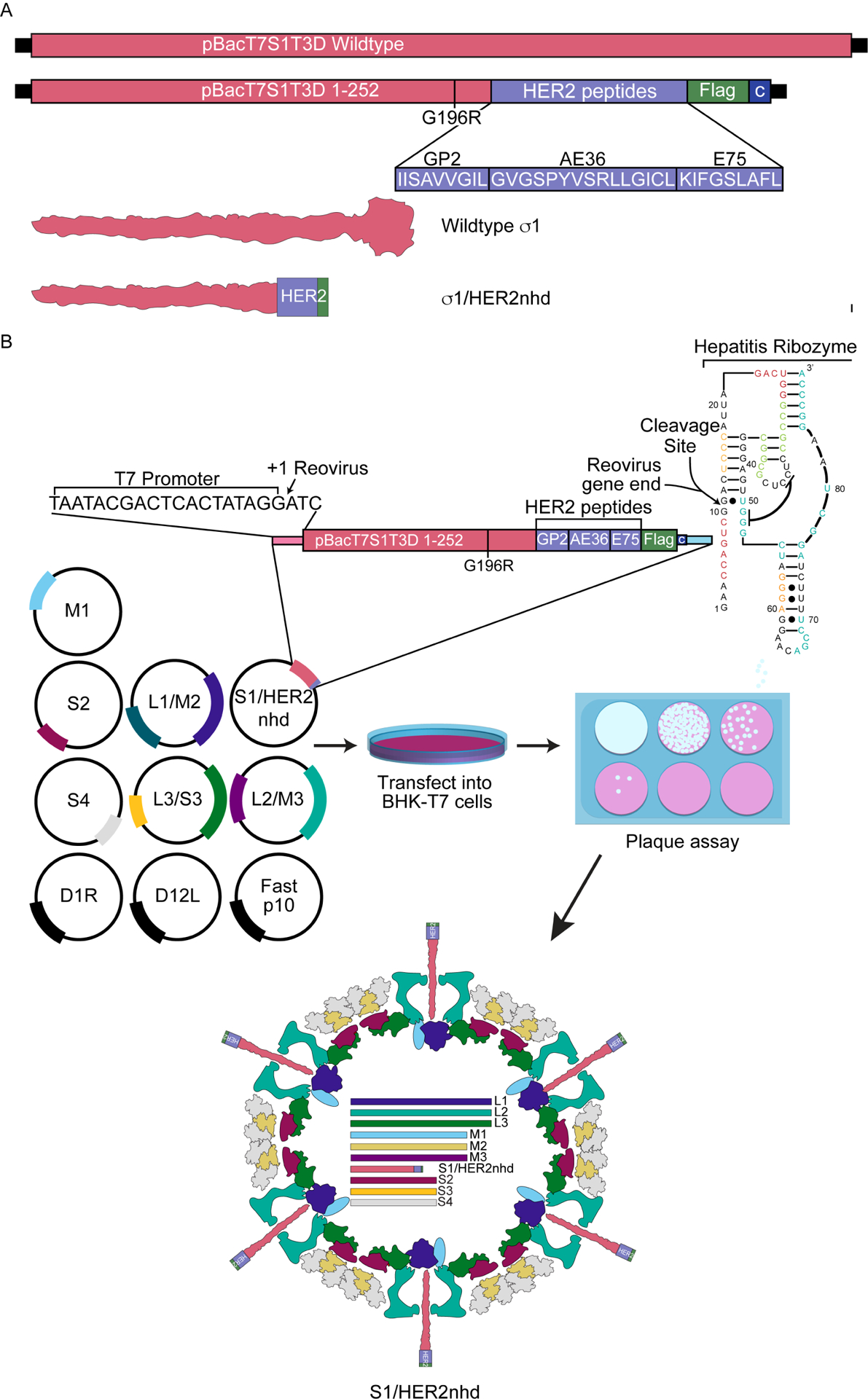
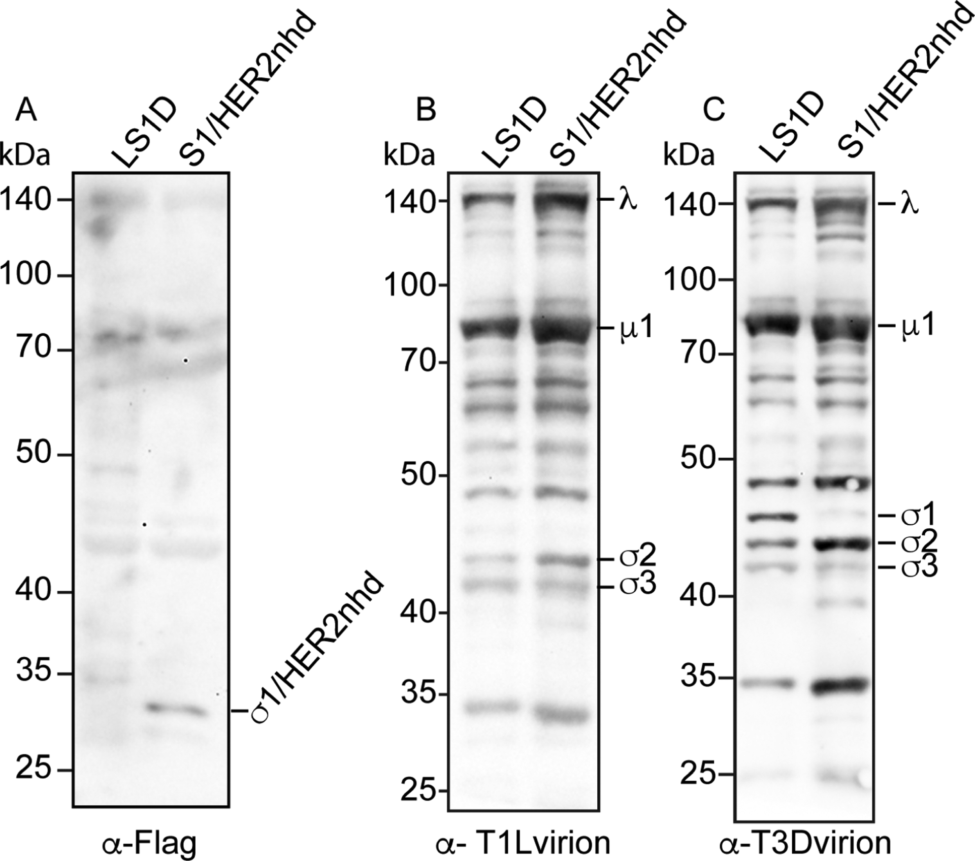
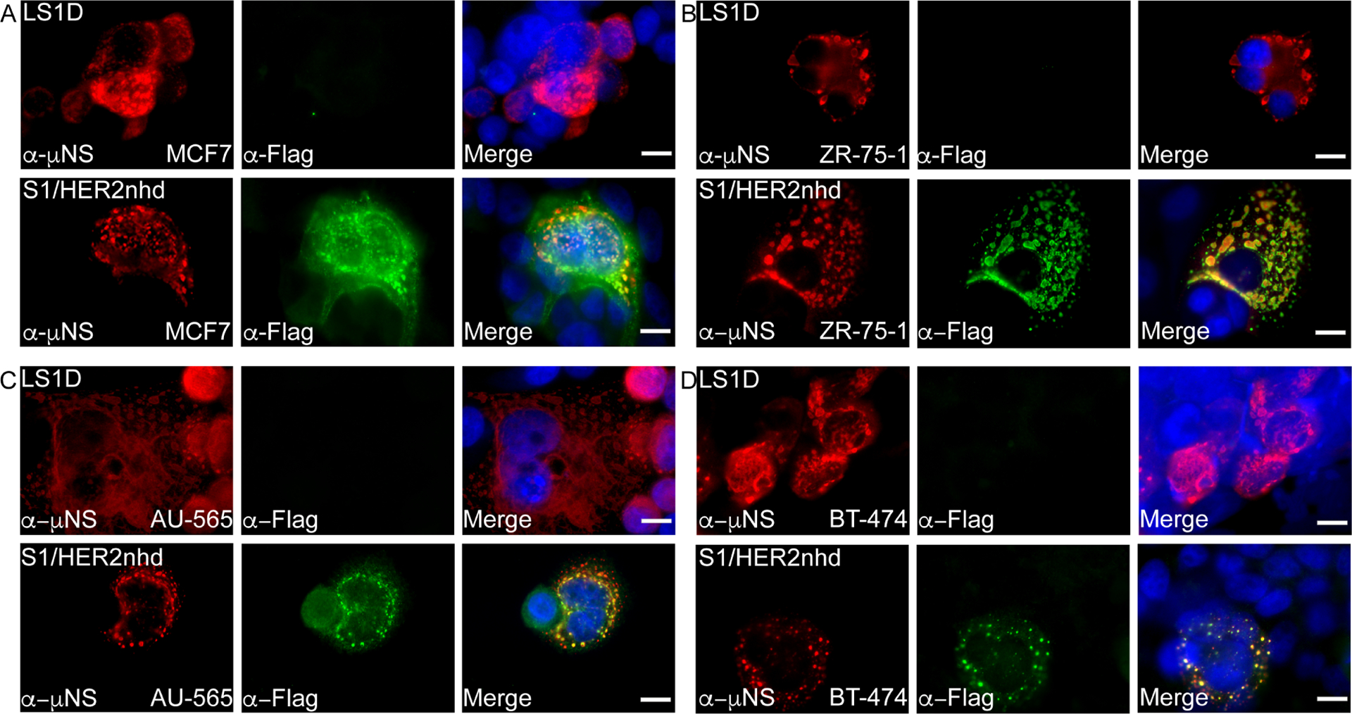
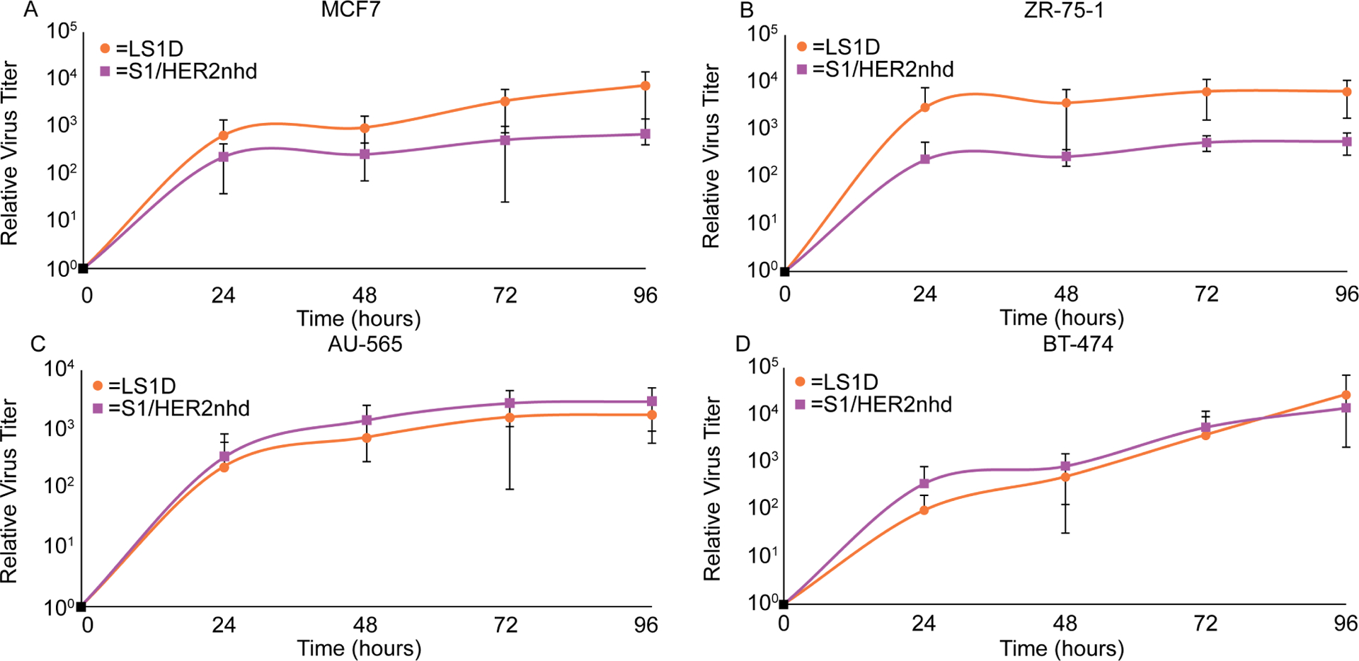
Mammalian orthoreovirus (MRV) is an oncolytic virus that has been tested in over 30 clinical trials. Increased clinical success has been achieved when MRV is used in combination with other onco-immunotherapies. This has led the field to explore the creation of recombinant MRVs which incorporate immunotherapeutic sequences into the virus genome. This work focuses on creation and characterization of a recombinant MRV, S1/HER2nhd, which encodes a truncated σ1 protein fused in frame with three human epidermal growth factor receptor 2 (HER2) peptides (E75, AE36, and GP2) known to induce HER2 specific CD8+ and CD4+ T cells. We show S1/HER2nhd expresses the σ1 fusion protein containing HER2 peptides in infected cells and on the virion, and infects, replicates in, and reduces survival of HER2+ breast cancer cells. The oncolytic properties of MRV combined with HER2 peptide expression holds potential as a vaccine to prevent recurrences of HER2 expressing cancers.

Keywords: Breast cancer; Cancer vaccine; HER2; Mammalian orthoreovirus; Oncolytic virus.


Conflict of interest statement

Declaration of competing interest The authors declare that they have no known competing financial interests or personal relationships that could have appeared to influence the work reported in this paper.

Figures

Fig 1.
Fig 1.. Design and generation of S1/HER2nhd.
A) Comparison of the WT pBacT7S1T3D with pBacT7S1/HER2nhd. pBacT7S1/HER2nhd contains WT Type 3 Dearing (T3D) S1 sequence until nt 767 (aa 252) with a nt substitution from 596 to 598 of GGG to CGA (aa G196R) followed by the coding sequence for HER2 peptides GP2, E75 and AE36, a Flag tag, a stop codon, and the C box, comprised of the last 124 nucleotides of S1 needed for S1 packaging. Beneath the plasmid construct illustration are the GP2, E75, and AE36 peptide sequences, and a schematic of the structure for σ1 and σ1/HER2nhd proteins. B) Reverse Genetics protocol used to generate S1/HER2nhd virus. WT T1L MRV plasmids pT7S2, pT7S4, pT7M1, pT7L1/M2, pT7L2/M3, and pT7L3/S3, along with pBacT7T3DS1/HER2nhd, and accessory plasmids D1R, D12L and Fast p10 were transfected into BHK-T7 cells and incubated at 37°C for 5 days. Samples were subjected to three freeze/thaw cycles and plaque assays were performed. Recombinant virus plaques were collected and passaged and RT-PCR products from viral RNA were sequenced to confirm S1/HER2nhd.
Fig 2.
Fig 2.. S1/HER2nhd genomic RNA and proteins are of expected size.
A) 1.88 × 10¹¹ virions of T1L, T3D, LS1D, and S1/HER2nhd were separated on SDS-PAGE and stained with 3X GelRed. B) 5 × 10¹⁰ virions of LS1D or S1/HER2nhd were separated on SDS-PAGE and stained with Coomassie Blue dye.
Fig 3.
Fig 3.. Flag antibodies recognize σ1/HER2nhd on MRV virions.
5 × 10¹⁰ virions of LS1D or S1/HER2nhd were separated on SDS-PAGE, before being transferred to nitrocellulose and stained with A) rabbit α-Flag polyclonal antibodies, B) rabbit α-TIL virion antisera, or C) rabbit α-T3D virion antisera, followed by HRP-conjugated α-rabbit secondary antibodies, exposure to chemiluminescent reagent, and image capture.
Fig 4.
Fig 4.. S1/HER2nhd stability upon passage.
Three plaques were picked after reverse genetics and passaged 10 times. RT-PCR products from RNA isolated from passage 3, 4, 6, 8 and 10 were sequenced. Numbers indicate nucleotide in the original σ1/HER2nhd before deletions. A) Plaque 1 sequence was stable through passage 6, but at passage 8 the boxed sequence changed from CAA to TAA, introducing a stop codon before the HER2 peptide sequence. B) Plaque 2 contained a partial deletion of the HER2 peptides starting at passage 4, maintaining the “IIS” sequence, but deleting the remaining HER2 peptide/Flag tag sequence and introducing a stop codon. C) Plaque 3 introduced the same stop codon as Plaque 1 in passage 3 which was fully established in the population by passage 4.
Fig 5.
Fig 5.. S1/HER2nhd and LS1D bind L929 cells with similar efficiency.
L929 cells were mock-infected or infected with A-C) 5 × 10¹⁰ particles of LS1D or S1/HER2nhd or D-F) MOI=1 LS1D or S1/HER2nhd. Cells were incubated with virus at 4°C for 1 hour, cell and supernatant fractions were collected and analyzed for viral titer using plaque assays. Percent bound virus is also shown (C, F). The means and standard deviation of three replicates are shown and the p-values, calculated using a 2-sided student T test, are as indicated.
Fig 6.
Fig 6.. S1/HER2nhd expresses HER2peptide/Flag fusion in infected cells.
BC cell lines A) MCF7 (HER2 0), B) ZR-75–1 (HER2 2+), C) AU-565 (HER2 3+) and D) BT-474 (HER2 3+) were infected with LS1D (top) or S1/HER2nhd (bottom) for 24 hours then subjected to immunofluorescent assay, staining with polyclonal α-μNS rabbit (red) and monoclonal α-Flag mouse (green) followed by Alexa 594-conjugated α-rabbit and Alexa 488-conjugated α-mouse secondary antibodies. Cells were counterstained with DAPI (blue) and merged images are shown. Scale Bar =10μm.
Fig 7.
Fig 7.. S1/HER2nhd and LS1D replicate to similar titers in BC cells.
A) MCF7, B) ZR-75–1, C) AU-565, and D) BT-474 cells were infected with LS1D or S1/HER2nhd at an MOI of 0.1. At indicated time points, samples were subjected to three freeze/thaw cycles and plaque assays on L929 cells. Samples from all timepoints were made relative to the average titer at time 0. The means and standard deviation from three experimental replicates are shown.
Fig 8.
Fig 8.. S1/HER2nhd reduces cell survival in BC cells.
A) MCF7, B) ZR-75–1, C) AU-565, and D) BT-474 cells were mock infected or infected at an MOI of 1 with LS1D or S1/HER2nhd. At the indicated time points CCK-8 reagent was added to manufactures specifications and analyzed. All values were made relative to mock infected average at that timepoint. The means and standard deviation of three replicates are shown and significant differences between samples are indicated. P-values calculated using 2-sided student T test are shown.

References

    1. Adair RA, Roulstone V, Scott KJ, Morgan R, Nuovo GJ, Fuller M, Beirne D, West EJ, Jennings VA, Rose A, Kyula J, Fraser S, Dave R, Anthoney DA, Merrick A, Prestwich R, Aldouri A, Donnelly O, Pandha H, Coffey M, Selby P, Vile R, Toogood G, Harrington K, Melcher AA, 2012. Cell carriage, delivery, and selective replication of an oncolytic virus in tumor in patients. Sci Transl Med 4, 138ra177. - PMC - PubMed
    1. Andreasson K, Tegerstedt K, Eriksson M, Curcio C, Cavallo F, Forni G, Dalianis T, Ramqvist T, 2009. Murine pneumotropic virus chimeric Her2/neu virus-like particles as prophylactic and therapeutic vaccines against Her2/neu expressing tumors. Int J Cancer 124, 150–156. - PubMed
    1. Andtbacka RH, Kaufman HL, Collichio F, Amatruda T, Senzer N, Chesney J, Delman KA, Spitler LE, Puzanov I, Agarwala SS, Milhem M, Cranmer L, Curti B, Lewis K, Ross M, Guthrie T, Linette GP, Daniels GA, Harrington K, Middleton MR, Miller WH, Zager JS, Ye Y, Yao B, Li A, Doleman S, VanderWalde A, Gansert J, Coffin RS, 2015. Talimogene Laherparepvec Improves Durable Response Rate in Patients With Advanced Melanoma. J Clin Oncol 33, 2780–2788. - PubMed
    1. Andtbacka RHI, Collichio F, Harrington KJ, Middleton MR, Downey G, Ӧhrling K, Kaufman HL, 2019. Final analyses of OPTiM: a randomized phase III trial of talimogene laherparepvec versus granulocyte-macrophage colony-stimulating factor in unresectable stage III-IV melanoma. J Immunother Cancer 7, 145. - PMC - PubMed
    1. Barton ES, Connolly JL, Forrest JC, Chappell JD, Dermody TS, 2001a. Utilization of sialic acid as a coreceptor enhances reovirus attachment by multistep adhesion strengthening. J Biol Chem 276, 2200–2211. - PubMed

Publication types