

USP13 regulates cell senescence through mediating MDM2 stability
- PMID: 37634814
- PMCID: PMC10807248
- DOI: 10.1016/j.lfs.2023.122044
USP13 regulates cell senescence through mediating MDM2 stability
Abstract
Aims: Lung aging results in altered lung function, reduced lung remodeling and regenerative capacity, and increased susceptibility to acute and chronic lung diseases. The molecular and physiological underlying mechanisms of lung aging remain unclear. Mounting evidence suggests that deubiquitinating enzymes (DUBs) play a critical role in tissue aging and diseases through regulation of cellular signaling pathways. Here we investigate the role of Ubiquitin-Specific Protease 13 (USP13) in cell senescence and lung aging and its underlying mechanisms.
Main methods: Protein levels of USP13 and MDM2 in lung tissues from aged and young mice were compared. Gene silencing and overexpression of USP13 in human cell lines were performed. MDM2 levels were examined by Quantitative Real-Time PCR and Western blotting analysis. The cell senescence levels of human cells were checked by the β-galactosidase staining.
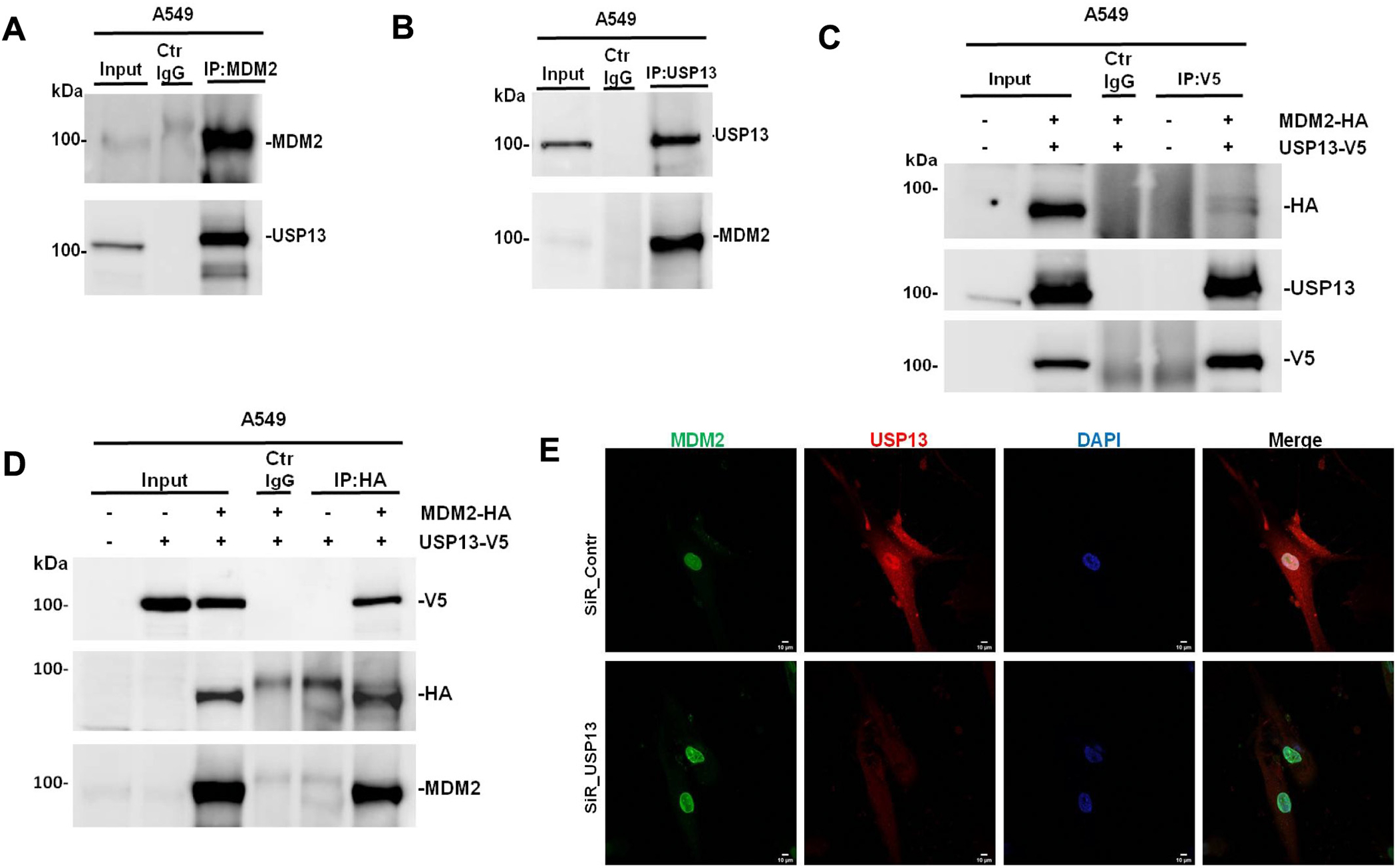
Key findings: Lung tissues from aged mice showed higher levels of USP13 compared to younger mice. We found a negative correlation between USP13 and MDM2 expression in lung tissues of aged mice. The increased protein levels of MDM2 were detected in lung tissues of USP13 deficient mice. Furthermore, overexpression of USP13 promoted cell senescence. Knockdown of USP13 increased MDM2 levels in lung cells, while overexpression of USP13 reduced it. The degradation of MDM2 caused by USP13 was prevented by the proteasome inhibitor MG132. Furthermore, we showed that USP13 targeted and reduced K63-linked polyubiquitination of MDM2. These results demonstrate that USP13 is involved in the aging signaling pathway in lungs through regulation of MDM2.
Keywords: Cell senescence; Deubiquitination; Lung aging; MDM2; USP13.
Copyright © 2023. Published by Elsevier Inc.
Conflict of interest statement
Declaration of competing interest The authors declare no competing interest.
Figures


Similar articles
-
The deubiquitinase USP13 stabilizes the anti-inflammatory receptor IL-1R8/Sigirr to suppress lung inflammation.EBioMedicine. 2019 Jul;45:553-562. doi: 10.1016/j.ebiom.2019.06.011. Epub 2019 Jun 14. EBioMedicine. 2019. PMID: 31204278 Free PMC article.
-
USP13 genetics and expression in a family with thyroid cancer.Endocrine. 2022 Aug;77(2):281-290. doi: 10.1007/s12020-022-03068-x. Epub 2022 May 18. Endocrine. 2022. PMID: 35583846 Free PMC article.
-
USP13 mediates PTEN to ameliorate osteoarthritis by restraining oxidative stress, apoptosis and inflammation via AKT-dependent manner.Biomed Pharmacother. 2021 Jan;133:111089. doi: 10.1016/j.biopha.2020.111089. Epub 2020 Dec 9. Biomed Pharmacother. 2021. PMID: 33378983
-
USP13: A therapeutic target for combating tumorigenesis and antitumor therapy resistance.Int J Biol Macromol. 2025 Apr;304(Pt 1):140608. doi: 10.1016/j.ijbiomac.2025.140608. Epub 2025 Feb 1. Int J Biol Macromol. 2025. PMID: 39900156 Review.
-
Regulatory Role of Ubiquitin Specific Protease-13 (USP13) in Misfolded Protein Clearance in Neurodegenerative Diseases.Neuroscience. 2021 Apr 15;460:161-166. doi: 10.1016/j.neuroscience.2021.02.004. Epub 2021 Feb 10. Neuroscience. 2021. PMID: 33577955 Review.
Cited by
-
Molecular Regulation of Transforming Growth Factor-β1-induced Thioredoxin-interacting Protein Ubiquitination and Proteasomal Degradation in Lung Fibroblasts: Implication in Pulmonary Fibrosis.J Respir Biol Transl Med. 2024 Mar;1(1):10002. doi: 10.35534/jrbtm.2024.10002. Epub 2024 Feb 1. J Respir Biol Transl Med. 2024. PMID: 38529321 Free PMC article.
-
Friend or foe? Reciprocal regulation between E3 ubiquitin ligases and deubiquitinases.Biochem Soc Trans. 2024 Feb 28;52(1):241-267. doi: 10.1042/BST20230454. Biochem Soc Trans. 2024. PMID: 38414432 Free PMC article. Review.
-
Matrix-free human lung organoids derived from induced pluripotent stem cells to model lung injury.Stem Cell Res Ther. 2024 Dec 18;15(1):468. doi: 10.1186/s13287-024-04106-3. Stem Cell Res Ther. 2024. PMID: 39696649 Free PMC article.
-
The mitochondrial enzyme pyruvate carboxylase restricts pancreatic β-cell senescence by blocking p53 activation.Proc Natl Acad Sci U S A. 2024 Oct 29;121(44):e2401218121. doi: 10.1073/pnas.2401218121. Epub 2024 Oct 22. Proc Natl Acad Sci U S A. 2024. PMID: 39436667 Free PMC article.
-
Immunoinflammation and post-translational modifications in the aging process.J Transl Med. 2025 Aug 14;23(1):910. doi: 10.1186/s12967-025-06892-7. J Transl Med. 2025. PMID: 40814109 Free PMC article. Review.
References
-
- Angelidis Ilias, Simon Lukas M., Fernandez Isis E., Strunz Maximilian, Mayr Christoph H., Greiffo Flavia R., Tsitsiridis George, Ansari Meshal, Graf Elisabeth, Strom Tim-Matthias, Nagendran Monica, Desai Tushar, Eickelberg Oliver, Mann Matthias, Theis Fabian J., Schiller Herbert B., An atlas of the aging lung mapped by single cell transcriptomics and deep tissue proteomics, Nat. Commun. 10 (1) (2019. Feb 27) 963, 10.1038/s41467-019-08831-9. - DOI - PMC - PubMed
-
- Jana Nihar Ranjan, Protein homeostasis and aging: role of ubiquitin protein ligases, Neurochem. Int. 60 (2012) (2012) 443–447. - PubMed
-
- Zhang Zhuo, Wang Hui, Mao Li, Agrawal Sudhir, Chen Xinbin, Zhang Ruiwen, MDM2 is a negative regulator of p21WAF1/CIP1, independent of p53, J. Biol. Chem. 279 (16) (2004. Apr 16) 16000–16006. - PubMed
-
- Marine J-C, Lozano G, Mdm2-mediated ubiquitylation: p53 and beyond, Cell Death Differ. 17 (2010) 93–102. - PubMed
MeSH terms
Substances
Grants and funding
LinkOut - more resources
Full Text Sources
Molecular Biology Databases
Research Materials
