

Radiological Study of Atlas Arch Defects with Meta-Analysis and a Proposed New Classification
- PMID: 37634902
- PMCID: PMC10622819
- DOI: 10.31616/asj.2023.0030
Radiological Study of Atlas Arch Defects with Meta-Analysis and a Proposed New Classification
Abstract
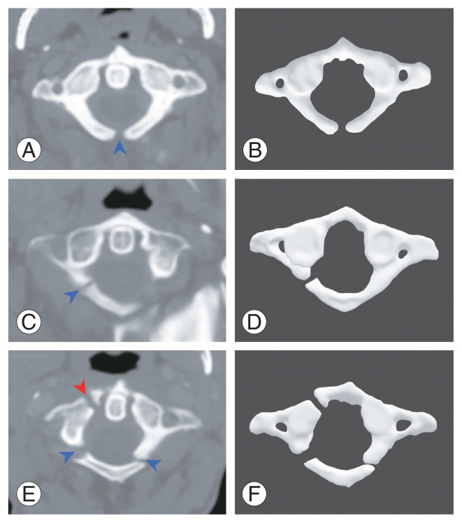
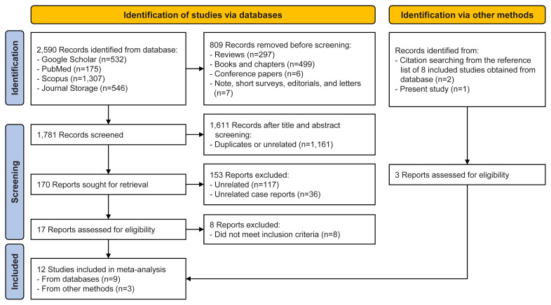
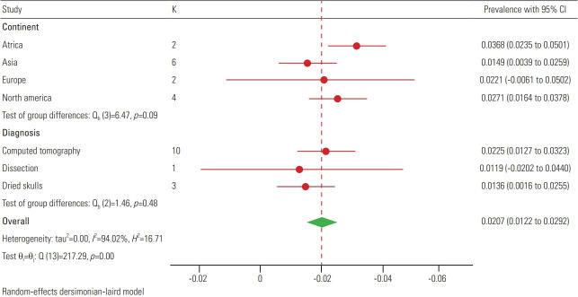
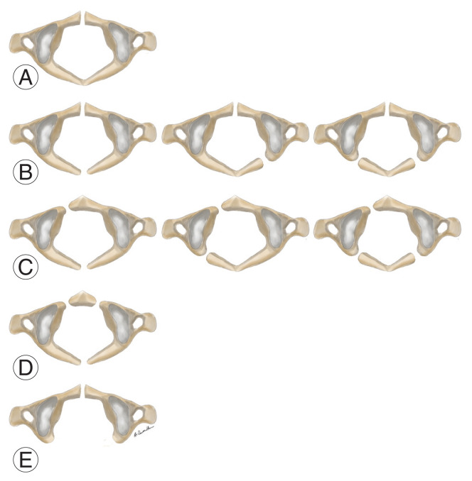
This study consists of a retrospective cohort study, a systematic review, and a meta-analysis which were separately conducted. This study aimed to investigate the prevalence of atlas arch defects, generate an evidence-based synthesis, and propose a common classification system for the anterior and combined atlas arch defects. Atlas arch defects are well-corticated gaps in the anterior or posterior arch of the atlas. When both arches are involved, it is known as a combined arch defect. Awareness of these defects is essential for avoiding complications during surgical procedures on the upper spine. The prevalence of arch defects was investigated in an open-access OPC-Radiomics (Radiomic Biomarkers in Oropharyngeal Carcinoma) dataset comprising 606 head and neck computed tomography scans from oropharyngeal cancer patients. A systematic review and meta-analysis were performed to generate prevalence estimates of atlas arch defects and propose a classification system for the anterior and combined atlas arch defects. The posterior arch defect was found in 20 patients (3.3%) out of the 606 patients investigated. The anterior arch defect was not observed in any patient, while a combined arch defect was observed in one patient (0.2%). A meta-analysis of 13,539 participants from 14 studies, including the present study, yielded a pooled-posterior arch defect prevalence of 2.07% (95% confidence interval [CI], 1.22%-2.92%). The prevalences of anterior and combined arch defects were 0.00% (95% CI, 0.00%-0.10%) and 0.14% (95% CI, 0.04%-0.25%), respectively. The anterior and combined arch defects were classified into five subtypes based on their morphology and frequency. The present study showed that atlas arch defects were present in approximately 2% of the general population. For future studies, larger sample sizes should be used for studying arch defects to avoid the small-study effect and to predict the prevalence accurately.
Keywords: Cervical atlas; Computed tomography; Meta-analysis; Systematic review.
Conflict of interest statement
The present study did not meet the criteria for ethical approval according to the self-assessment form issued by the Mahidol University Central Institutional Review Board (MU-CIRB). Data sharing will be available upon reasonable request to the corresponding author. This research did not receive any specific grant from funding agencies in the public, commercial, or not-for-profit sectors. Furthermore, the authors declare no conflicts of interest in this manuscript.
No potential conflict of interest relevant to this article was reported.
Figures
References
-
- Garg A, Gaikwad SB, Gupta V, Mishra NK, Kale SS, Singh J. Bipartite atlas with os odontoideum: case report. Spine (Phila Pa 1976) 2004;29:E35–8. - PubMed
-
- Garg S, Agarwal R, Goyal D. Bipartite atlas: a rare entity, a study of its incidence in North Indians. Ann Int Med Dent Res. 2018;4:AT01–3.
-
- Hu Y, Ma W, Xu R. Transoral osteosynthesis C1 as a function-preserving option in the treatment of bipartite atlas deformity: a case report. Spine (Phila Pa 1976) 2009;34:E418–21. - PubMed
-
- Sibbou K, Chalh O, Zamani O, Hoummadi A, El Fenni J. Split atlas mimicking a fracture. Joint Bone Spine. 2022;89:105386. - PubMed
-
- Tachibana A, Imabayashi H, Yato Y, Nakamichi K, Asazuma T, Nemoto K. Torticollis of a specific C1 dislocation with split atlas. Spine (Phila Pa 1976) 2010;35:E672–5. - PubMed
Grants and funding
LinkOut - more resources
Full Text Sources
