. 2024 Feb;40(2):157-170.
doi: 10.1007/s12264-023-01102-0. Epub 2023 Aug 27.

Temporal Unfolding of Racial Ingroup Bias in Neural Responses to Perceived Dynamic Pain in Others

Affiliations

Temporal Unfolding of Racial Ingroup Bias in Neural Responses to Perceived Dynamic Pain in Others

Chenyu Pang et al. Neurosci Bull. 2024 Feb.

Abstract

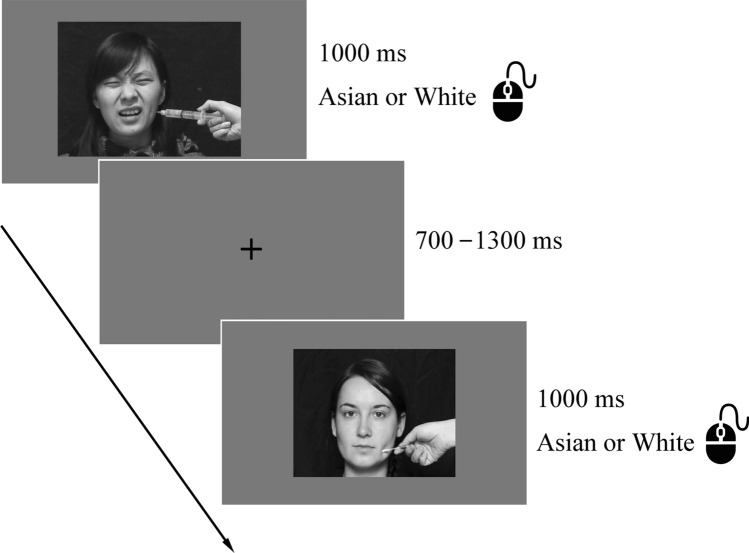
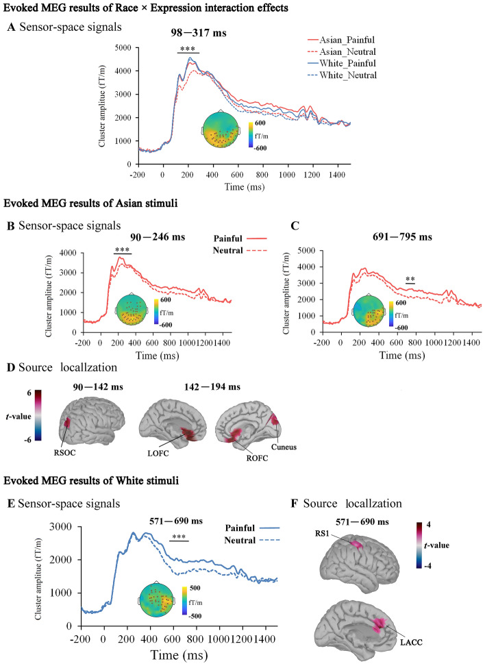
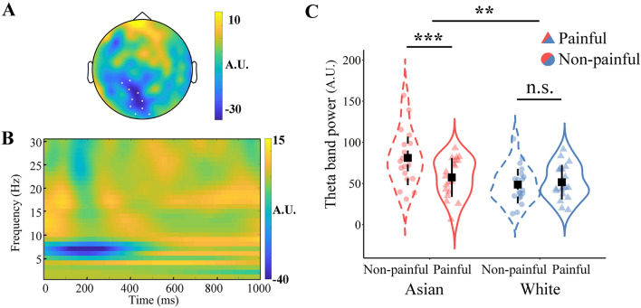
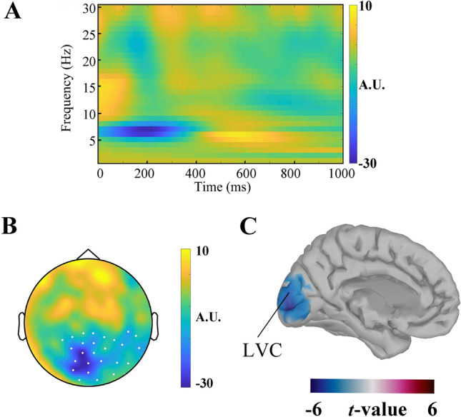
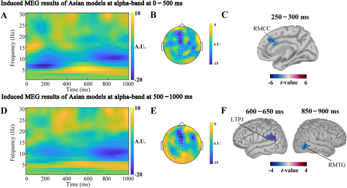
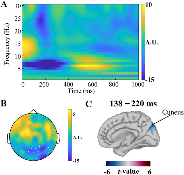
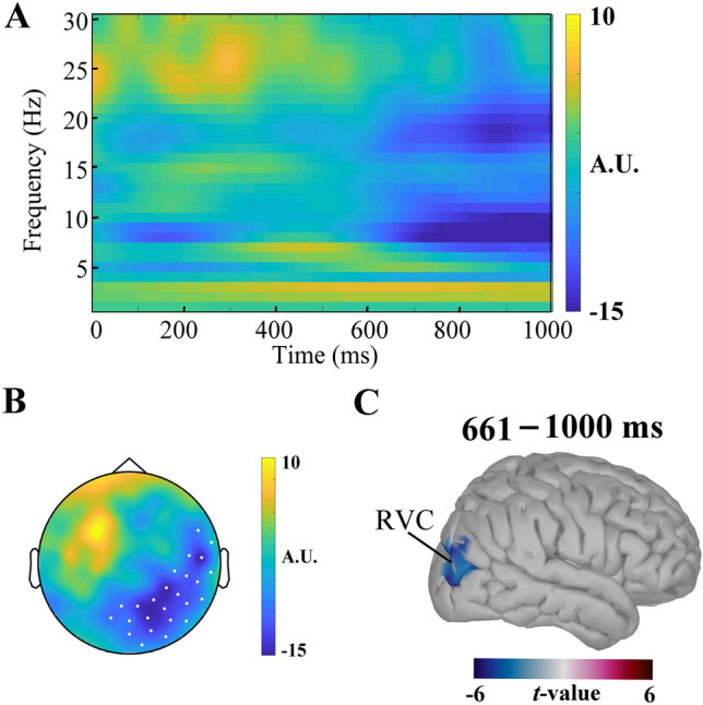
In this study, we investigated how empathic neural responses unfold over time in different empathy networks when viewing same-race and other-race individuals in dynamic painful conditions. We recorded magnetoencephalography signals from Chinese adults when viewing video clips showing a dynamic painful (or non-painful) stimulation to Asian and White models' faces to trigger painful (or neutral) expressions. We found that perceived dynamic pain in Asian models modulated neural activities in the visual cortex at 100 ms-200 ms, in the orbitofrontal and subgenual anterior cingulate cortices at 150 ms-200 ms, in the anterior cingulate cortex around 250 ms-350 ms, and in the temporoparietal junction and middle temporal gyrus around 600 ms after video onset. Perceived dynamic pain in White models modulated activities in the visual, anterior cingulate, and primary sensory cortices after 500 ms. Our findings unraveled earlier dynamic activities in multiple neural circuits in response to same-race (vs other-race) individuals in dynamic painful situations.

Keywords: Cingulate cortex; Dynamic pain; Empathy; Magnetoencephalography; Race.


Conflict of interest statement

The authors declare no competing financial interests.

Figures

Fig. 1
Fig. 1
Illustration of the procedure during MEG recording. Participants were asked to respond to the racial identities of models in video clips any time after the onset of a video clip .
Fig. 2
Fig. 2
Results of time-domain sensor-space and source space MEG signals. A Illustration of the time window and sensor locations in which sensor-space MEG signals showed significant Race × Expression interaction effects. The topography shows the location of sensors showing significant interaction effects. B Sensor-space MEG signals in response to Asian models. The topography shows the location of sensors that showed significantly greater responses to painful (vs non-painful) stimuli to Asian models. C Sensor-space MEG signals in response to Asian models. D Source brain regions in which neural responses were significantly stronger to painful (vs non-painful) stimuli to Asian models. E Sensor-space MEG signals in response to White models. The topography shows the location of sensors that showed significantly greater responses to painful (vs non-painful) stimuli to White models. F Source brain regions in which neural responses were significantly stronger to painful (vs non-painful) stimuli to White models. ROFC, right orbitofrontal cortex; LOFC, left orbitofrontal cortex; RSC, right sensory cortex; LMCC, left midcingulate cortex. Repeated-measures ANOVA, n = 21, **P <0.01, ***P <0.001.
Fig. 3
Fig. 3
Interaction effects on induced theta-band responses to others’ pain. A Topography of sensor-space signals that showed significant Race × Stimulus valence interaction effects (defined by the contrast of Asian faces (Painful-Non-painful) vs White faces (Painful-Non-painful)) at 0 ms–336 ms. White dots indicate sensors in the significant cluster. B The mean values of theta power across time averaged from all the sensors in the significant cluster. C Race × Stimulus valence interaction effects on theta power. Mean theta power in each condition (black squares) across all participants, the mean theta power of each participant (a dot or a triangle), and the one SD from the mean of each group (lower and higher whiskers). Significant interaction effects were identified using a cluster threshold of P <0.05 for sensor space two-tailed and P <0.005 for source-space one-tailed. Repeated-measures ANOVA, n = 21, *P <0.05, **P <0.01, ***P <0.001, n.s., no significant difference.
Fig. 4
Fig. 4
Theta responses to Asian models in painful and non-painful conditions. A The mean values of theta power across time and sensors in the significant cluster. B Topography of sensor-space signals that showed significant modulations by painful vs non-painful stimuli applied to Asian models at 0 ms–285 ms. White dots indicate sensors in the significant cluster. C The brain region contributing to the effect of painful vs non-painful stimuli on sensor-space signals. The empathy effect of MEG signals was identified using a cluster threshold of P <0.05 for sensor space two-tailed and P <0.005 for source-space one-tailed. LVC, left visual cortex.
Fig. 5
Fig. 5
Alpha-band responses to painful vs non-painful stimuli to Asian models. A The mean values of alpha power across time and sensors in the significant cluster at 0 ms–500 ms. B Topography of sensor-space signals that showed significant modulation of alpha responses by painful vs non-painful stimuli to Asian models at 141 ms–361 ms. White dots indicate sensors in the significant cluster. C Source brain regions contributing to the effect of painful vs non-painful stimuli on alpha power. D The mean values of alpha power across time and sensors in the significant cluster at 500 ms–1000 ms. E Topography of sensor-space signals that showed significant modulation of alpha responses by painful vs non-painful stimuli to Asian models at 607 ms–1000 ms. White dots indicate sensors in the significant cluster. F Source brain regions contributing to the effect of painful vs non-painful stimuli on alpha power. MCC, midcingulate cortex; LTPJ, left temporoparietal junction; RMTG, right middle temporal gyrus; LVC, left visual cortex. The effect of painful vs non-painful stimuli was identified using a cluster threshold of P <0.05 for sensor space two-tailed and P <0.005 for source-space one-tailed, n = 21.
Fig. 6
Fig. 6
Beta band responses to painful vs non-painful stimuli to Asian models. A The mean value of beta power across time and sensors in the significant cluster at 0 ms–500 ms. B Topography of sensor-space signals that showed significant modulations of beta responses by painful vs non-painful stimuli applied to Asian models at 138 ms–220 ms. White dots indicate sensors in the significant cluster. C The brain region contributing to the effect of painful vs non-painful stimuli on beta power in response to Asian models.
Fig. 7
Fig. 7
Alpha band responses to painful vs non-painful stimuli to White models. A The mean values of beta power across time and sensors in the significant cluster at 500 ms–1000 ms. B Topography of sensor-space signals that showed significant modulation of beta responses by painful vs non-painful stimuli applied to White models at 661 ms–1000 ms. White dots indicate sensors in the significant clusters. C The brain region contributing to the effect of painful vs non-painful stimuli on alpha power in response to White models. RVC, right visual cortex.
Fig. 8
Fig. 8
Relationships between IAT D scores and neural responses to painful (vs non-painful) stimuli. A larger IAT D score is associated with a smaller evoked sensor-level response to painful (vs non-painful) stimuli to White models.

Similar articles

Cited by

References

    1. Brown SL, Cialdini RB. Functional motives and functional consequences of prosocial behavior. In: The Oxford Handbook of Prosocial Behavior. Oxford University Press, 2015: 346–361.
    1. Chen J. Empathy for distress in humans and rodents. Neurosci Bull. 2018;34:216–236. doi: 10.1007/s12264-017-0135-0. - DOI - PMC - PubMed
    1. Decety J, Bartal IB, Uzefovsky F, Knafo-Noam A. Empathy as a driver of prosocial behaviour: Highly conserved neurobehavioural mechanisms across species. Philos Trans R Soc Lond B Biol Sci. 2016;371:20150077. doi: 10.1098/rstb.2015.0077. - DOI - PMC - PubMed
    1. Johnson JD, Simmons CH, Jordav A, MacLean L, Taddei J, Thomas D, et al. Rodney King and O.J. revisited: The impact of race and defendant empathy induction on judicial decisions. J Appl Social Pyschol. 2002;32:1208–1223. doi: 10.1111/j.1559-1816.2002.tb01432.x. - DOI
    1. Luo S, Li B, Ma Y, Zhang W, Rao Y, Han S. Oxytocin receptor gene and racial ingroup bias in empathy-related brain activity. Neuroimage. 2015;110:22–31. doi: 10.1016/j.neuroimage.2015.01.042. - DOI - PubMed

LinkOut - more resources