. 2023 Aug 27;21(1):301.
doi: 10.1186/s12951-023-02028-5.

A simple and robust nanosystem for photoacoustic imaging of bladder cancer based on α5β1-targeted gold nanorods

Affiliations

A simple and robust nanosystem for photoacoustic imaging of bladder cancer based on α5β1-targeted gold nanorods

Massimo Alfano et al. J Nanobiotechnology. .

Abstract

Background: Early detection and removal of bladder cancer in patients is crucial to prevent tumor recurrence and progression. Because current imaging techniques may fail to detect small lesions of in situ carcinomas, patients with bladder cancer often relapse after initial diagnosis, thereby requiring frequent follow-up and treatments.

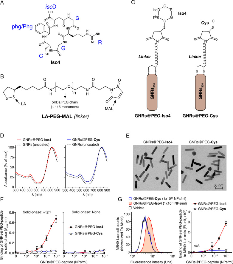
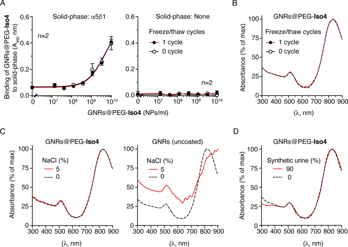
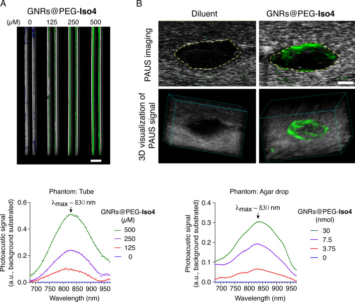
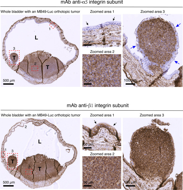
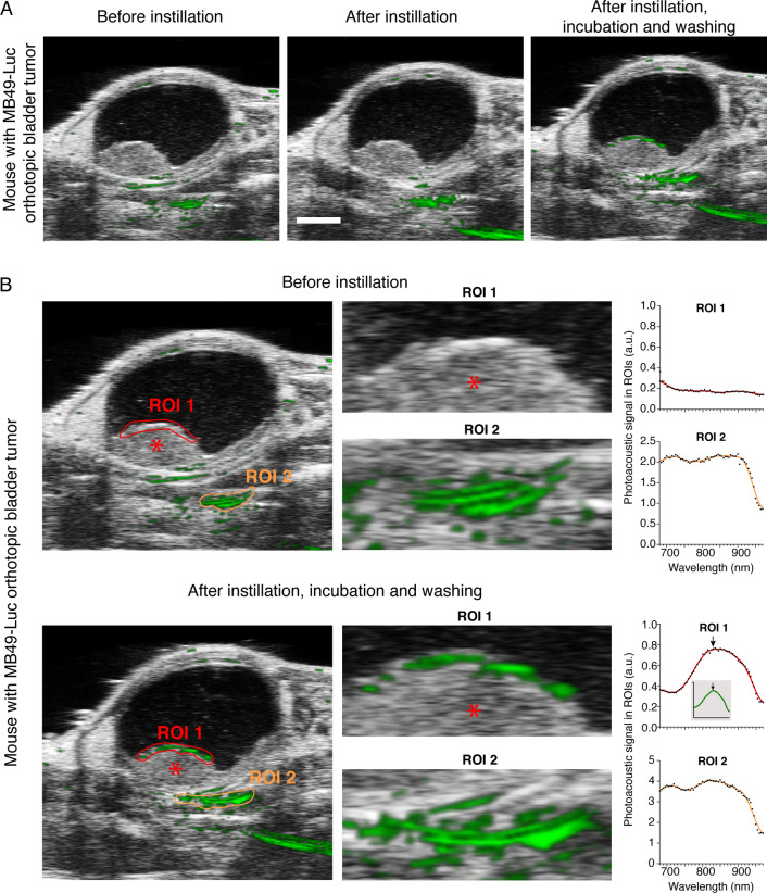
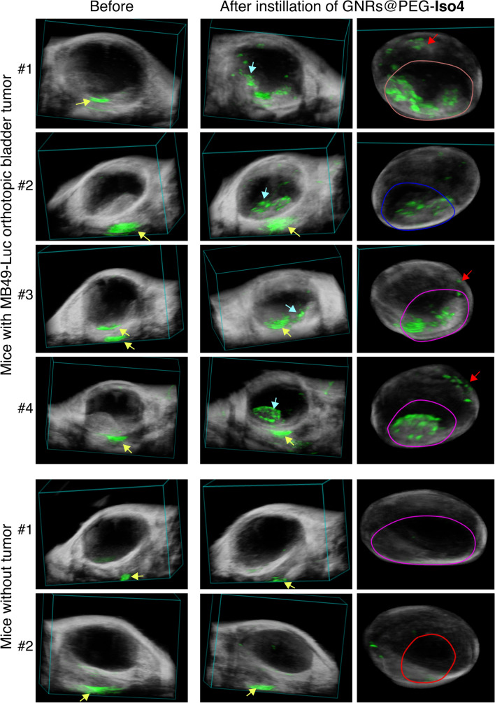
Results: In an attempt to obtain a sensitive and high-resolution imaging modality for bladder cancer, we have developed a photoacoustic imaging approach based on the use of PEGylated gold nanorods (GNRs) as a contrast agent, functionalized with the peptide cyclic [CphgisoDGRG] (Iso4), a selective ligand of α5β1 integrin expressed by bladder cancer cells. This product (called GNRs@PEG-Iso4) was produced by a simple two-step procedure based on GNRs activation with lipoic acid-polyethyleneglycol(PEG-5KDa)-maleimide and functionalization with peptide Iso4. Biochemical and biological studies showed that GNRs@PEG-Iso4 can efficiently recognize purified integrin α5β1 and α5β1-positive bladder cancer cells. GNRs@PEG-Iso4 was stable and did not aggregate in urine or in 5% sodium chloride, or after freeze/thaw cycles or prolonged exposure to 55 °C, and, even more importantly, do not settle after instillation into the bladder. Intravesical instillation of GNRs@PEG-Iso4 into mice bearing orthotopic MB49-Luc bladder tumors, followed by photoacoustic imaging, efficiently detected small cancer lesions. The binding to tumor lesions was competed by a neutralizing anti-α5β1 integrin antibody; furthermore, no binding was observed to healthy bladders (α5β1-negative), pointing to a specific targeting mechanism.

Conclusion: GNRs@PEG-Iso4 represents a simple and robust contrast agent for photoacoustic imaging and diagnosis of small bladder cancer lesions.

Keywords: Bladder cancer; Gold nanorods; IsoDGR motif; Photoacoustic imaging; α5β1 integrin.


Conflict of interest statement

M.A., A.C., and F.C. are inventors of patents regarding GNRs functionalized with peptide Iso4.

Figures

Fig. 1
Fig. 1
Schematic representation and characterization of GNRs@PEG-Iso4 and GNRs@PEG-Cys. Representation of the structures of: A head-to-tail cyclized peptide Iso4, B lipoic acid-PEG-maleimide heterobifunctional cross-linker (LA-PEG-MAL) containing a PEG chain of 5KDa, C and gold nanorods (GNRs) functionalized with peptide Iso4 (GNRs@PEG-Iso4) or Cys (GNRs@PEG-Cys) via LA-PEG-MAL. D UV-IR absorption spectra of the GNRs@PEG-Iso4 and GNRs@PEG-Cys. The dotted line corresponds to the uncoated gold nanorods (GNRs). E Representative microphotographs of GNRs@PEG-Iso4 and GNRs@PEG-Cys, as determined by transmission electron microscopy (TEM). F Binding of GNRs@PEG-Iso4 and GNRs@PEG-Cys to plates coated with or without α5β1-integrin. The binding of nanoparticles was detected with an anti-PEG rat antibody followed by HRP-labelled goat anti-rat antiserum. Mean ± SE of technical duplicates. G Binding of GNRs@PEG-Iso4 and GNRs@PEG-Cys to bladder cancer MB49-Luc cells as determined by FACS. MB49-Luc cells in suspension were incubated with the indicated amounts of nanoparticles for 1 h on ice. After washing, the cells were incubated with an anti-PEG antibody (0.5 h on ice) followed by a FITC-labelled secondary antibody (0.5 h in ice). The bound fluorescence was quantified by flow cytometry analysis. Representative FACS plots (left) and dose-dependent binding curves (right) (dots, mean ± SE of technical triplicates) are shown
Fig. 2
Fig. 2
Stability studies of GNRs@PEG-Iso4. A Effect of one freeze–thaw cycle on the binding properties of GNRs@PEG-Iso4 to α5β1. The binding of GNRs@PEG-Iso4 to plates coated with or without α5β1 integrin before (0 cycle) and after one freezing (-80 °C) and thawing cycle (1 cycle), as detected with an anti-PEG rat antibody followed by HRP-labeled goat anti-rat antiserum, is shown. Mean ± SE of technical duplicates. Note that the freeze–thaw cycle does not affect the α5β1-binding properties of GNRs@PEG-Iso4. B-D Stability studies of GNRs@PEG-Iso4 and GNRs nanoparticles, performed by UV-IR absorption analysis of: i) nanoparticles before (0 cycle) and after one freezing (-80 °C) and thawing cycle (1 cycle) (B), ii) nanoparticles mixed with or without sodium chloride (5% NaCl, final concentration) (C), and iii) nanoparticles mixed with synthetic urine (90% urine, final concentration) (D). Note that the addition of sodium chloride caused a dramatic change in the UV-IR absorption spectrum of GNRs, but not of GNRs@PEG-Iso4, suggesting that the latter compound is protected from aggregation induced by high salt concentration
Fig. 3
Fig. 3
In vitro PA and US imaging of GNRs@PEG-Iso4. A Representative 2D PA and US image of capillary tubes filled with the indicated amount of GNRs@PEG-Iso4 (upper), and quantification of PA signal (lower). Grayscale, co-registered US signal; Green, PA signal of NPs. Bar, 1 mm. B Representative 2D and 3D PA and US images (upper and middle panels, respectively) of an agar drop with or without GNRs@PEG-Iso4 (30 µl nanogold, 30 nmol Au, ~ 1.16 × 1011 NPs), and quantification of PA signal (lower). The yellow dotted line in the upper marks the boundary between agar drop and the slime gel. Grayscale, co-registered US signal; Green: PA signal. Bar, 1 mm. Note that GNRs accumulated mainly at the periphery of the drop during the polymerization of the agar/GNRs mixture
Fig. 4
Fig. 4
Expression of α5- and β1-integrin subunit in the MB49-Luc bladder cancer model. Representative immunohistochemistry photomicrographs of the expression of α5- and β1-integrin subunit in different areas of the bladder of a tumor-bearing mice, 11 days after intravesical instillation of MB49-Luc cells. This mouse developed two MB49-Luc tumors (T) with different size. L, lumen of the urinary bladder; Red dashed rectangle: zoomed areas. Zoomed area 1: healthy bladder, black arrows indicate the urothelial cells. Zoomed area 2: Inner MB49-Luc tumor. Zoomed area 3: Small tumor. The small tumor, but not the big one, is almost completely covered by 2–5 layers of α5-negative urothelial cells (blue arrows). Scale bar is shown in each panel. Immunostaining was performed as described previously [15]. See also Additional File 1: Fig. S7
Fig. 5
Fig. 5
In vivo 2D PA and US images of an orthotopic MB49-Luc tumor before and after administration of GNRs@PEG-Iso4. MB49-Luc cells were implanted orthotopically in the bladder of a mouse. After 14 days, the bladder was imaged by PA and US analysis before and immediately after intravesical instillation of GNRs@PEG-Iso4 (26 nmol Au, ~ 1 × 1011 NPs), and after 15 min of incubation and bladder washing. A A representative axial 2D PA and US images of the entire bladder according to the indicated time of analysis. Grayscale, co-registered US signal; Green, PA signal Bar, 2 mm. B Zoomed images of Panel A and PA spectra in the selected ROIs according to the indicated time of analysis. Asterisk, Tumor; Red and Orange features delineate the ROIs drawn on the apical part of the tumor (ROI 1) and outside the bladder (ROI 2), respectively. Note that the PA spectrum detected in the tumor after installation of GNRs@PEG-Iso4, but not that of the PA signal observed outside the bladder, is very similar to the spectrum obtained with the same nanoparticles dispersed in agar drops (inset). Arrows, λmax: ~ 830 nm
Fig. 6
Fig. 6
GNRs@PEG-Iso4 binds to orthotopic MB49-Luc bladder tumors but not to healthy bladder tissue. Representative axial 2D PA and US images of the bladder from tumor-bearing mice (n = 4 mice) or from healthy control mice (n = 2 mice) before and after the instillation of GNRs@PEG-Iso4 (26 nmol Au, ~ 1 × 1011 NPs), followed by incubation and bladder washing. Tumor-bearing mice were PA and US imaged 11–14 days after MB49-Luc cell implantation. Grayscale, co-registered US signal; Green, PA signal. Cyan arrows, PA signal generated by GNRs@PEG-Iso4. Yellow arrows, PA signal independent of GNRs@PEG-Iso4. Bar, 2 mm
Fig. 7.
Fig. 7.
3D PA and US images of bladders from MB49-Luc tumor-bearing mice or healthy control mice before and after instillation of GNRs@PEG-Iso4. Representative 3D visualization (left and middle panels) and 3D reconstruction (right panels) of the PA and US signal of the bladders of mice of Fig. 6. Grayscale, co-registered ultrasound signal; Green, PA signal. Cyan arrows, specific signal of GNRs@PEG-Iso4. Yellow arrows, nonspecific signal recorded outside the bladder present before administration of GNRs@PEG-Iso4. Note the almost complete absence of PA signal inside the bladder in control animals. Red arrows indicate small spots of PA signal likely corresponding to small bladder cancer lesions (< 0.5 mm) that are undetectable by standard US echography

References

    1. Ferlay J, Soerjomataram I, Dikshit R, Eser S, Mathers C, Rebelo M, et al. Cancer incidence and mortality worldwide: sources, methods and major patterns in GLOBOCAN 2012. Int J Cancer. 2015;136(5):E359–E386. doi: 10.1002/ijc.29210. - DOI - PubMed
    1. Burger M, Catto JW, Dalbagni G, Grossman HB, Herr H, Karakiewicz P, et al. Epidemiology and risk factors of urothelial bladder cancer. Eur Urol. 2013;63(2):234–241. doi: 10.1016/j.eururo.2012.07.033. - DOI - PubMed
    1. https://uroweb.org/guidelines/non-muscle-invasive-bladder-cancer. Accessed 10 Aug 2023.
    1. Ozden E, Turgut AT, Turkolmez K, Resorlu B, Safak M. Effect of bladder carcinoma location on detection rates by ultrasonography and computed tomography. Urology. 2007;69(5):889–892. doi: 10.1016/j.urology.2007.01.077. - DOI - PubMed
    1. Babjuk M, Burger M, Comperat EM, Gontero P, Mostafid AH, Palou J, et al. European association of urology guidelines on non-muscle-invasive bladder cancer (TaT1 and carcinoma in situ)—2019 update. Eur Urol. 2019;76(5):639–657. doi: 10.1016/j.eururo.2019.08.016. - DOI - PubMed

LinkOut - more resources