

Targeted delivery of a PD-1-blocking scFv by CD133-specific CAR-T cells using nonviral Sleeping Beauty transposition shows enhanced antitumour efficacy for advanced hepatocellular carcinoma
- PMID: 37635247
- PMCID: PMC10464109
- DOI: 10.1186/s12916-023-03016-0
Targeted delivery of a PD-1-blocking scFv by CD133-specific CAR-T cells using nonviral Sleeping Beauty transposition shows enhanced antitumour efficacy for advanced hepatocellular carcinoma
Abstract
Background: CD133 is considered a marker for cancer stem cells (CSCs) in several types of tumours, including hepatocellular carcinoma (HCC). Chimeric antigen receptor-specific T (CAR-T) cells targeting CD133-positive CSCs have emerged as a tool for the clinical treatment of HCC, but immunogenicity, the high cost of clinical-grade recombinant viral vectors and potential insertional mutagenesis limit their clinical application.
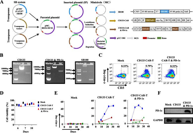
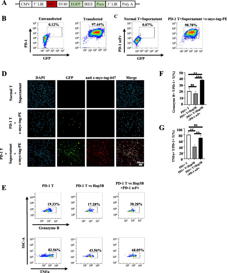
Methods: CD133-specific CAR-T cells secreting PD-1 blocking scFv (CD133 CAR-T and PD-1 s cells) were constructed using a sleeping beauty transposon system from minicircle technology, and the antitumour efficacy of CD133 CAR-T and PD-1 s cells was analysed in vitro and in vivo.
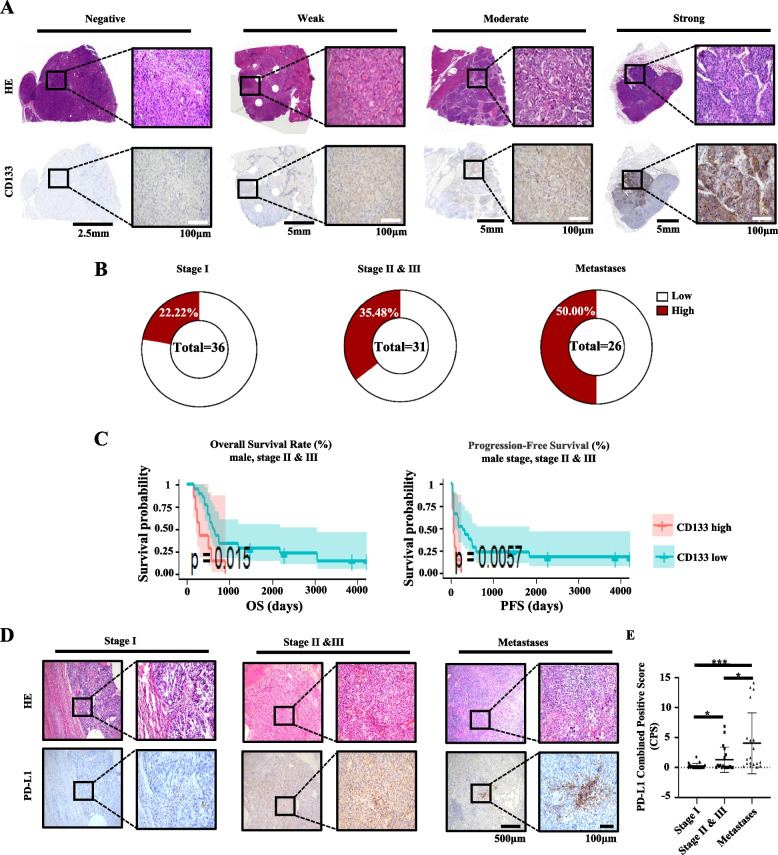
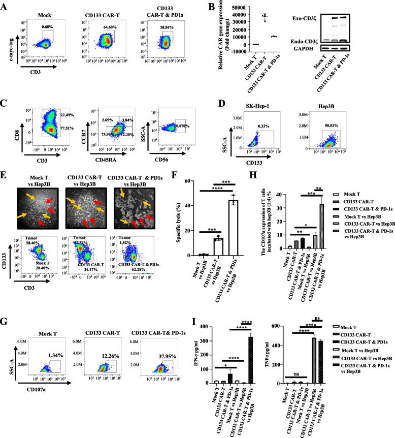
Results: A univariate analysis showed that CD133 expression in male patients at the late stage (II and III) was significantly associated with worse progression-free survival (PFS) (P = 0.0057) and overall survival (OS) (P = 0.015), and a multivariate analysis showed a trend toward worse OS (P = 0.041). Male patients with advanced HCC exhibited an approximately 20-fold higher PD-L1 combined positive score (CPS) compared with those with HCC at an early stage. We successfully generated CD133 CAR-T and PD-1 s cells that could secrete PD-1 blocking scFv based on a sleeping beauty system involving minicircle vectors. CD133 CAR-T and PD-1 s cells exhibited significant antitumour activity against HCC in vitro and in xenograft mouse models. Thus, CD133 CAR-T and PD-1 s cells may be a therapeutically tractable strategy for targeting CD133-positive CSCs in male patients with advanced HCC.
Conclusions: Our study provides a nonviral strategy for constructing CAR-T cells that could also secrete checkpoint blockade inhibitors based on a Sleeping Beauty system from minicircle vectors and revealed a potential benefit of this strategy for male patients with advanced HCC and high CD133 expression (median immunohistochemistry score > 2.284).
Keywords: CAR-T; CD133; Hepatocellular carcinoma; Minicircle; PD-1; Sleeping Beauty system.
© 2023. BioMed Central Ltd., part of Springer Nature.
Conflict of interest statement
The authors declare that they have no competing interests.
Figures


Similar articles
-
Nonviral mcDNA-mediated bispecific CAR T cells kill tumor cells in an experimental mouse model of hepatocellular carcinoma.BMC Cancer. 2022 Jul 25;22(1):814. doi: 10.1186/s12885-022-09861-1. BMC Cancer. 2022. PMID: 35879685 Free PMC article.
-
Nucleofection with Plasmid DNA for CRISPR/Cas9-Mediated Inactivation of Programmed Cell Death Protein 1 in CD133-Specific CAR T Cells.Hum Gene Ther. 2019 Apr;30(4):446-458. doi: 10.1089/hum.2017.234. Epub 2018 Apr 27. Hum Gene Ther. 2019. PMID: 29706119
-
CAR T Cells Generated Using Sleeping Beauty Transposon Vectors and Expanded with an EBV-Transformed Lymphoblastoid Cell Line Display Antitumor Activity In Vitro and In Vivo.Hum Gene Ther. 2019 Apr;30(4):511-522. doi: 10.1089/hum.2018.218. Hum Gene Ther. 2019. PMID: 30793967
-
Chimeric antigen receptor-engineered T-cell therapy for liver cancer.Hepatobiliary Pancreat Dis Int. 2018 Aug;17(4):301-309. doi: 10.1016/j.hbpd.2018.05.005. Epub 2018 May 24. Hepatobiliary Pancreat Dis Int. 2018. PMID: 29861325 Review.
-
Neoantigen Specific T Cells Derived From T Cell-Derived Induced Pluripotent Stem Cells for the Treatment of Hepatocellular Carcinoma: Potential and Challenges.Front Immunol. 2021 May 13;12:690565. doi: 10.3389/fimmu.2021.690565. eCollection 2021. Front Immunol. 2021. PMID: 34054880 Free PMC article. Review.
Cited by
-
Recent advancements in improving the efficacy and safety of chimeric antigen receptor (CAR)-T cell therapy for hepatocellular carcinoma.Naunyn Schmiedebergs Arch Pharmacol. 2025 Feb;398(2):1433-1446. doi: 10.1007/s00210-024-03443-7. Epub 2024 Sep 24. Naunyn Schmiedebergs Arch Pharmacol. 2025. PMID: 39316087 Review.
-
Chimeric Antigen Receptor T Cell Therapy for Hepatocellular Carcinoma: Where Do We Stand?Int J Mol Sci. 2024 Feb 23;25(5):2631. doi: 10.3390/ijms25052631. Int J Mol Sci. 2024. PMID: 38473878 Free PMC article. Review.
-
The synergistic immunotherapeutic impact of engineered CAR-T cells with PD-1 blockade in lymphomas and solid tumors: a systematic review.Front Immunol. 2024 May 10;15:1389971. doi: 10.3389/fimmu.2024.1389971. eCollection 2024. Front Immunol. 2024. PMID: 38799440 Free PMC article.
-
CAR-NK cells for gastrointestinal cancer immunotherapy: from bench to bedside.Mol Cancer. 2024 Oct 23;23(1):237. doi: 10.1186/s12943-024-02151-3. Mol Cancer. 2024. PMID: 39443938 Free PMC article. Review.
-
Exploration of the role of immune cells and cell therapy in hepatocellular carcinoma.Front Immunol. 2025 Apr 16;16:1569150. doi: 10.3389/fimmu.2025.1569150. eCollection 2025. Front Immunol. 2025. PMID: 40308592 Free PMC article. Review.
References
-
- Sung H, Ferlay J, Siegel RL, Laversanne M, Soerjomataram I, Jemal A, et al. Global cancer statistics 2020: GLOBOCAN estimates of incidence and mortality worldwide for 36 cancers in 185 Countries. CA Cancer J Clin. 2021;71(3):209–249. - PubMed
-
- Zhu AX, Finn RS, Edeline J, Cattan S, Ogasawara S, Palmer D, et al. Pembrolizumab in patients with advanced hepatocellular carcinoma previously treated with sorafenib (KEYNOTE-224): a non-randomised, open-label phase 2 trial. Lancet Oncol. 2018;19(7):940–952. - PubMed
Publication types
MeSH terms
Substances
LinkOut - more resources
Full Text Sources
Medical
Research Materials
