A comparative study of in vitro air-liquid interface culture models of the human airway epithelium evaluating cellular heterogeneity and gene expression at single cell resolution
- PMID: 37635251
- PMCID: PMC10464153
- DOI: 10.1186/s12931-023-02514-2
A comparative study of in vitro air-liquid interface culture models of the human airway epithelium evaluating cellular heterogeneity and gene expression at single cell resolution
Abstract
Background: The airway epithelium is composed of diverse cell types with specialized functions that mediate homeostasis and protect against respiratory pathogens. Human airway epithelial (HAE) cultures at air-liquid interface are a physiologically relevant in vitro model of this heterogeneous tissue and have enabled numerous studies of airway disease. HAE cultures are classically derived from primary epithelial cells, the relatively limited passage capacity of which can limit experimental methods and study designs. BCi-NS1.1, a previously described and widely used basal cell line engineered to express hTERT, exhibits extended passage lifespan while retaining the capacity for differentiation to HAE. However, gene expression and innate immune function in BCi-NS1.1-derived versus primary-derived HAE cultures have not been fully characterized.
Methods: BCi-NS1.1-derived HAE cultures (n = 3 independent differentiations) and primary-derived HAE cultures (n = 3 distinct donors) were characterized by immunofluorescence and single cell RNA-Seq (scRNA-Seq). Innate immune functions were evaluated in response to interferon stimulation and to infection with viral and bacterial respiratory pathogens.
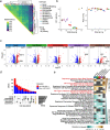
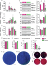
Results: We confirm at high resolution that BCi-NS1.1- and primary-derived HAE cultures are largely similar in morphology, cell type composition, and overall gene expression patterns. While we observed cell-type specific expression differences of several interferon stimulated genes in BCi-NS1.1-derived HAE cultures, we did not observe significant differences in susceptibility to infection with influenza A virus and Staphylococcus aureus.
Conclusions: Taken together, our results further support BCi-NS1.1-derived HAE cultures as a valuable tool for the study of airway infectious disease.
Keywords: Airway epithelium; Air–liquid interface epithelial culture; Innate immunity; Respiratory infection; Single cell transcriptomics.
© 2023. BioMed Central Ltd., part of Springer Nature.
Conflict of interest statement
The authors declare no competing interests.
Figures


Update of
-
A comparative study of in vitro air-liquid interface culture models of the human airway epithelium evaluating cellular heterogeneity and gene expression at single cell resolution.bioRxiv [Preprint]. 2023 Feb 28:2023.02.27.530299. doi: 10.1101/2023.02.27.530299. bioRxiv. 2023. Update in: Respir Res. 2023 Aug 28;24(1):213. doi: 10.1186/s12931-023-02514-2. PMID: 36909601 Free PMC article. Updated. Preprint.
Similar articles
-
A comparative study of in vitro air-liquid interface culture models of the human airway epithelium evaluating cellular heterogeneity and gene expression at single cell resolution.bioRxiv [Preprint]. 2023 Feb 28:2023.02.27.530299. doi: 10.1101/2023.02.27.530299. bioRxiv. 2023. Update in: Respir Res. 2023 Aug 28;24(1):213. doi: 10.1186/s12931-023-02514-2. PMID: 36909601 Free PMC article. Updated. Preprint.
-
Membrane-Tethered Mucin 1 Is Stimulated by Interferon and Virus Infection in Multiple Cell Types and Inhibits Influenza A Virus Infection in Human Airway Epithelium.mBio. 2022 Aug 30;13(4):e0105522. doi: 10.1128/mbio.01055-22. Epub 2022 Jun 14. mBio. 2022. PMID: 35699372 Free PMC article.
-
Long-term differentiating primary human airway epithelial cell cultures: how far are we?Cell Commun Signal. 2021 May 27;19(1):63. doi: 10.1186/s12964-021-00740-z. Cell Commun Signal. 2021. PMID: 34044844 Free PMC article. Review.
-
Long-Term Modeling of SARS-CoV-2 Infection of In Vitro Cultured Polarized Human Airway Epithelium.mBio. 2020 Nov 6;11(6):e02852-20. doi: 10.1128/mBio.02852-20. mBio. 2020. PMID: 33158999 Free PMC article.
-
Development, validation and implementation of an in vitro model for the study of metabolic and immune function in normal and inflamed human colonic epithelium.Dan Med J. 2015 Jan;62(1):B4973. Dan Med J. 2015. PMID: 25557335 Review.
Cited by
-
KLF4 promotes a KRT13+ hillock-like state in squamous lung cancer.bioRxiv [Preprint]. 2025 Mar 13:2025.03.10.641898. doi: 10.1101/2025.03.10.641898. bioRxiv. 2025. PMID: 40161723 Free PMC article. Preprint.
-
Challenges in current inhalable tobacco toxicity assessment models: A narrative review.Tob Induc Dis. 2024 Jun 10;22. doi: 10.18332/tid/188197. eCollection 2024. Tob Induc Dis. 2024. PMID: 38860150 Free PMC article. Review.
-
MHC class II proteins mediate sialic acid independent entry of human and avian H2N2 influenza A viruses.Nat Microbiol. 2024 Oct;9(10):2626-2641. doi: 10.1038/s41564-024-01771-1. Epub 2024 Jul 15. Nat Microbiol. 2024. PMID: 39009691
-
3D human tissue models and microphysiological systems for HIV and related comorbidities.Trends Biotechnol. 2024 May;42(5):526-543. doi: 10.1016/j.tibtech.2023.10.008. Epub 2023 Dec 8. Trends Biotechnol. 2024. PMID: 38071144 Free PMC article. Review.
-
Nasal Mucociliary Epithelial Cell Culture Models for Studying Viral Infections.Methods Mol Biol. 2025;2890:237-252. doi: 10.1007/978-1-0716-4326-6_13. Methods Mol Biol. 2025. PMID: 39890731
References
MeSH terms
Substances
Grants and funding
LinkOut - more resources
Full Text Sources
Molecular Biology Databases

