Review
. 2023 Sep 14;43(10):1063-1090.
doi: 10.1093/asj/sjad173.

The Role of Calcium Hydroxylapatite (Radiesse) as a Regenerative Aesthetic Treatment: A Narrative Review

Review

The Role of Calcium Hydroxylapatite (Radiesse) as a Regenerative Aesthetic Treatment: A Narrative Review

Shino Bay Aguilera et al. Aesthet Surg J. .

Abstract

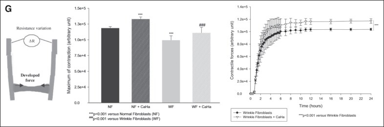
For decades, a wide variety of natural and synthetic materials have been used to augment human tissue to improve aesthetic outcomes. Dermal fillers are some of the most widely used aesthetic treatments throughout the body. Initially, the primary function of dermal fillers was to restore depleted volume. As biomaterial research has advanced, however, a variety of biostimulatory fillers have become staples in aesthetic medicine. Such fillers often contain a carrying vehicle and a biostimulatory material that induces de novo synthesis of major structural components of the extracellular matrix. One such filler, Radiesse (Merz Aesthetics, Raleigh, NC), is composed of calcium hydroxylapatite microspheres suspended in a carboxymethylcellulose gel. In addition to immediate volumization, Radiesse treatment results in increases of collagen, elastin, vasculature, proteoglycans, and fibroblast populations via a cell-biomaterial-mediated interaction. When injected, Radiesse acts as a cell scaffold and clinically manifests as immediate restoration of depleted volume, improvements in skin quality and appearance, and regeneration of endogenous extracellular matrices. This narrative review contextualizes Radiesse as a regenerative aesthetic treatment, summarizes its unique use cases, reviews its rheological, material, and regenerative properties, and hypothesizes future combination treatments in the age of regenerative aesthetics.


Figures

Figure 1.
Figure 1.
CaHA-R filler characterization data. (A) Scanning electron microscopy images of CaHA-R show uniformly sized, smooth, and defect-free microspheres. (B) Antibacterial properties of different filler types showing near-zero Staphylococci adherence to CaHA-R. (C) Elastic moduli and (D) Complex viscosity of different filler types measured at 0.7 Hz. CaHA, calcium hydroxylapatite; CaHA-R, calcium hydroxylapatite (Radiesse; Merz Aesthetics, Raleigh, NC); HA, hyaluronic acid. Juvederm Ultra Plus XC, Juvederm Ultra (Allergan, an AbbVie Compay, Chicago, IL), Belotero Balance (Merz Aesthetics, Raleigh, NC), Bellafill (polymethylmethacrylate; Suneva Medical, San Diego, CA), Macroplastique (polydimethylsiloxane/polyvinylpyrrolidone; Laborie Medical Technologies Corporation, Portsmouth, NH). Figure 1B is reproduced with permission from Wang et al (2022).  Figure 1C, D is reproduced with permission from Lorenc et al (2018).
Figure 2.
Figure 2.
Mechanobiology of aging. (A) Loss of tension and (B) loss of contact are proposed as 2 driving mechanobiological factors of aging. (C) Confocal images of α-actin–stained fibroblasts (brown) cultured with intact collagen (left) and degraded collagen (right) show differences in cellular morphologies. (D) Young (18- to 29-year-old) fibroblasts and old (80+-year-old) fibroblasts show different morphologies and are correlated with less organized and misaligned collagen fibers, resulting in aged fibroblasts having significantly less mechanoreceptor activation. (E) Healthy (sun protected) and damaged (sun damaged) fibroblasts show changes in morphology. Damaged fibroblasts have less contact with collagen, are less active, and are significantly more elongated than healthy fibroblasts. Figure 2A, D is reproduced with permission from Varani et al (2006).  Figure 2B, C, E is reproduced with permission from Varani et al (2004).
Figure 2.
Figure 2.
Mechanobiology of aging. (A) Loss of tension and (B) loss of contact are proposed as 2 driving mechanobiological factors of aging. (C) Confocal images of α-actin–stained fibroblasts (brown) cultured with intact collagen (left) and degraded collagen (right) show differences in cellular morphologies. (D) Young (18- to 29-year-old) fibroblasts and old (80+-year-old) fibroblasts show different morphologies and are correlated with less organized and misaligned collagen fibers, resulting in aged fibroblasts having significantly less mechanoreceptor activation. (E) Healthy (sun protected) and damaged (sun damaged) fibroblasts show changes in morphology. Damaged fibroblasts have less contact with collagen, are less active, and are significantly more elongated than healthy fibroblasts. Figure 2A, D is reproduced with permission from Varani et al (2006).  Figure 2B, C, E is reproduced with permission from Varani et al (2004).
Figure 3.
Figure 3.
Regenerative mechanism of action of CaHA-R. (A) Cell culture studies demonstrated dose-independent cell viability demonstrating excellent biocompatibility of CaHA-R. (B) The “stretch” mechanism of action hypothesized by Wang et al, which states that: (1) a hydrogel filler is shown as preferentially localizing in areas containing more highly fragmented collagen fibers, since these regions may be more accommodating; (2) this results in stretching of existing collagen fibers (curved lines), which is sensed by nearby fibroblasts through cell surface receptors such as integrins. In response, fibroblasts become morphologically stretched (3) and activated to produce extracellular matrix components, suggesting the volumetric displacement from the carboxymethylcellulose carrier gel may contribute to regeneration. (C) Collagen staining (blue) and regression analysis showing relationship between microsphere density and collagen regeneration. Green circles denote single microspheres; yellow circle denotes microsphere grouping; black triangluation denotes the distance calculation between microspheres. (D) Hematoxylin and eosin stains showing a capsule of stretched endogenous fibroblasts (denoted with *) encapsulating a CaHA microsphere surrounded by collagen fibers (Cf) and not in contact with macrophages (Mp). 1 m = 1 month post injection. (E) Clinical study showing the dilution-dependent regeneration of collagen I and III, thus highlighting the relevance of having space between microspheres to enable cell-biomaterial interactions. (F) Confocal microscopy images of de novo collagen I fibers arising from and connecting to neighboring CaHA microspheres, resulting in (G) highly aligned and dense collagen networks. (H) The working mechanism of action of CaHA-R: fibroblasts encounter CaHA microspheres and undergo direct mechanotransduction (activation of mechanoreceptors and stretching) that results in extracellular matrix protein synthesis. CaHA, calcium hydroxylapatite; CaHA-R, calcium hydroxylapatite (Radiesse; Merz Aesthetics, Raleigh, NC); FB, fibroblasts; KC, keratinocytes. Figure 3A is reproduced with permission from Wollina et al (2018).  Figure 3B is reproduced with permission from Wang et al (2007).  Figure 3D is reproduced with permission from Kim (2019).  Figure 3E is reproduced with permission from Casabona and Pereira (2017).
Figure 3.
Figure 3.
Regenerative mechanism of action of CaHA-R. (A) Cell culture studies demonstrated dose-independent cell viability demonstrating excellent biocompatibility of CaHA-R. (B) The “stretch” mechanism of action hypothesized by Wang et al, which states that: (1) a hydrogel filler is shown as preferentially localizing in areas containing more highly fragmented collagen fibers, since these regions may be more accommodating; (2) this results in stretching of existing collagen fibers (curved lines), which is sensed by nearby fibroblasts through cell surface receptors such as integrins. In response, fibroblasts become morphologically stretched (3) and activated to produce extracellular matrix components, suggesting the volumetric displacement from the carboxymethylcellulose carrier gel may contribute to regeneration. (C) Collagen staining (blue) and regression analysis showing relationship between microsphere density and collagen regeneration. Green circles denote single microspheres; yellow circle denotes microsphere grouping; black triangluation denotes the distance calculation between microspheres. (D) Hematoxylin and eosin stains showing a capsule of stretched endogenous fibroblasts (denoted with *) encapsulating a CaHA microsphere surrounded by collagen fibers (Cf) and not in contact with macrophages (Mp). 1 m = 1 month post injection. (E) Clinical study showing the dilution-dependent regeneration of collagen I and III, thus highlighting the relevance of having space between microspheres to enable cell-biomaterial interactions. (F) Confocal microscopy images of de novo collagen I fibers arising from and connecting to neighboring CaHA microspheres, resulting in (G) highly aligned and dense collagen networks. (H) The working mechanism of action of CaHA-R: fibroblasts encounter CaHA microspheres and undergo direct mechanotransduction (activation of mechanoreceptors and stretching) that results in extracellular matrix protein synthesis. CaHA, calcium hydroxylapatite; CaHA-R, calcium hydroxylapatite (Radiesse; Merz Aesthetics, Raleigh, NC); FB, fibroblasts; KC, keratinocytes. Figure 3A is reproduced with permission from Wollina et al (2018).  Figure 3B is reproduced with permission from Wang et al (2007).  Figure 3D is reproduced with permission from Kim (2019).  Figure 3E is reproduced with permission from Casabona and Pereira (2017).
Figure 4.
Figure 4.
CaHA-R regenerates different components of the skin. (A, B) Evidence of collagen turnover reveals the formation of new collagen after 2 months in vivo. (C) Histologic stains show significant increases in elastin. (D) Histologic stains show significant increases in proteoglycans (top), and rete ride depth (bottom) after treatment with CaHA. (E, F) Split-arm comparison of Sculptra (Galderma, Lausanne, Switzerland) and CaHA-R shows similar rates of collagen formation and higher staining intensity for elastin fibers with CaHA-R. (G) Anti-CD31 antibody staining revealing an abundance of neovasculature in the deep dermis, 1 month postinjection of CaHA. CaHA, calcium hydroxylapatite; CaHA-R, calcium hydroxylapatite (Radisesse; Merz Aesthetics, Raleigh, NC); PLLA, poly-L-lactic acid. Figure 4A, B is reproduced with permission from Zerbinati and Calligaro (2018).  Figure 4C, D is reproduced with permission from González and Goldberg (2019).  Figure 4E, F is reproduced with permission from Mazzuco et al (2022).  Figure 4G is reproduced with permission from Shalak OV, Satygo EA, Deev RV, Presnyakov EV, The Effectiveness of Radiesse in Dental Practice for Prevention and Non-Surgical Treatment of Gum Recession. Her North-West State Med Univ Named II Mechnikov. 2023, Volume 14, Issue 4, pages 43-52; use permitted under the Creative Commons Attribution License CC BY-NC-ND 4.0 (https://creativecommons.org/licenses/by-nc-nd/4.0/).
Figure 4.
Figure 4.
CaHA-R regenerates different components of the skin. (A, B) Evidence of collagen turnover reveals the formation of new collagen after 2 months in vivo. (C) Histologic stains show significant increases in elastin. (D) Histologic stains show significant increases in proteoglycans (top), and rete ride depth (bottom) after treatment with CaHA. (E, F) Split-arm comparison of Sculptra (Galderma, Lausanne, Switzerland) and CaHA-R shows similar rates of collagen formation and higher staining intensity for elastin fibers with CaHA-R. (G) Anti-CD31 antibody staining revealing an abundance of neovasculature in the deep dermis, 1 month postinjection of CaHA. CaHA, calcium hydroxylapatite; CaHA-R, calcium hydroxylapatite (Radisesse; Merz Aesthetics, Raleigh, NC); PLLA, poly-L-lactic acid. Figure 4A, B is reproduced with permission from Zerbinati and Calligaro (2018).  Figure 4C, D is reproduced with permission from González and Goldberg (2019).  Figure 4E, F is reproduced with permission from Mazzuco et al (2022).  Figure 4G is reproduced with permission from Shalak OV, Satygo EA, Deev RV, Presnyakov EV, The Effectiveness of Radiesse in Dental Practice for Prevention and Non-Surgical Treatment of Gum Recession. Her North-West State Med Univ Named II Mechnikov. 2023, Volume 14, Issue 4, pages 43-52; use permitted under the Creative Commons Attribution License CC BY-NC-ND 4.0 (https://creativecommons.org/licenses/by-nc-nd/4.0/).
Figure 5.
Figure 5.
CaHA-R can restore the structure and function of skin. (A) Results from a clinical study administering multiple layers of CaHA-R show significant reduction in wrinkles (left), significant reduction in keratinization (middle), and significant improvements in discoloration (right). (B) Cutometric analysis of CaHA-R-treated skin shows improvements in skin mechanical properties in the thighs, abdomen, and brachial zone. (C-F) Ultrasound analysis shows significant increases in the thickness of the skin and the subcutis 3 and 6 weeks after a single CaHA-R treatment. (G) In vitro study measuring fibroblast contractile force (left) in normal or wrinkled fibroblasts with CaHA revealed the restoration of contractile forces in wrinkled fibroblasts to the level of normal fibroblasts and the attainment of supraphysiological contractile forces in normal fibroblasts treated with CaHA. The changes in contractile force were consistent throughout the duration of the study. CaHA, calcium hydroxylapatite; CaHA-R, calcium hydroxylapatite (Radiesse; Merz Aesthetics, Raleigh, NC); NF, normal fibroblast; WF, wrinkle fibroblast. Figure 5A is reproduced with permission from Kim (2019).  Figure 5B is reproduced with permission from Wasylkowski (2015).  Figure 5F, G is reproduced with permission from Courderot-Masuyer C, Robin S, Tauzin H, Humbert P. Evaluation of Lifting and Antiwrinkle Effects of Calcium Hydroxylapatite Filler In Vitro Quantification of Contractile Forces of Human Wrinkle and Normal Aged Fibroblasts Treated with Calcium Hydroxylapatite. J Cosmet Dermatol. 2016, Volume 15, Issue 3, pages 260-268, with permission from John Wiley & Sons (https://onlinelibrary.wiley.com).
Figure 5.
Figure 5.
CaHA-R can restore the structure and function of skin. (A) Results from a clinical study administering multiple layers of CaHA-R show significant reduction in wrinkles (left), significant reduction in keratinization (middle), and significant improvements in discoloration (right). (B) Cutometric analysis of CaHA-R-treated skin shows improvements in skin mechanical properties in the thighs, abdomen, and brachial zone. (C-F) Ultrasound analysis shows significant increases in the thickness of the skin and the subcutis 3 and 6 weeks after a single CaHA-R treatment. (G) In vitro study measuring fibroblast contractile force (left) in normal or wrinkled fibroblasts with CaHA revealed the restoration of contractile forces in wrinkled fibroblasts to the level of normal fibroblasts and the attainment of supraphysiological contractile forces in normal fibroblasts treated with CaHA. The changes in contractile force were consistent throughout the duration of the study. CaHA, calcium hydroxylapatite; CaHA-R, calcium hydroxylapatite (Radiesse; Merz Aesthetics, Raleigh, NC); NF, normal fibroblast; WF, wrinkle fibroblast. Figure 5A is reproduced with permission from Kim (2019).  Figure 5B is reproduced with permission from Wasylkowski (2015).  Figure 5F, G is reproduced with permission from Courderot-Masuyer C, Robin S, Tauzin H, Humbert P. Evaluation of Lifting and Antiwrinkle Effects of Calcium Hydroxylapatite Filler In Vitro Quantification of Contractile Forces of Human Wrinkle and Normal Aged Fibroblasts Treated with Calcium Hydroxylapatite. J Cosmet Dermatol. 2016, Volume 15, Issue 3, pages 260-268, with permission from John Wiley & Sons (https://onlinelibrary.wiley.com).
Figure 6.
Figure 6.
Immunological response to CaHA-R. (A) Scant inflammatory cell activation both 1 and 6 months after CaHA-R treatment, despite the increase in collagen staining intensity (blue). (B) Evidence of eventual macrophage-mediated breakdown of CaHA microspheres. (C) Hematoxylin and eosin stains of the same magnification from a CaHA-R-treated and PLLA-SCA-treated arm show profound differences in immune cell infiltration at injection site. (D) Proposed gradient on particle morphology and its role in regulating inflammation. The smoother and more homogeneous a particle, the less inflammatory. (E) Scanning electron microscopy images of CaHA-R microspheres and (F) PLLA-SCA flakes reveal dramatic differences in size, shape, surface irregularities, and phagocytosable materials. (G) Significant differences in proinflammatory cytokine release from M1 macrophages cultured with CaHA and PLLA, demonstrating the minimal inflammatory potential of CaHA. CaHA, calcium hydroxylapatite; CaHA-R, calcium hydroxylapatite (Radiesse; Merz Aesthetics, Raleigh, NC); MIP1a, macrophage inflammatory protein 1α; PLLA, poly-L-lactic acid; PLLA-SCA, poly-L-lactic acid (Sculptra; Galderma, Lausanne, Switzerland); TNFRII, tumor necrosis factor receptor II. Figure 6A is reproduced with permission from Marmur et al (2004).  Figure 6C is reproduced with permission from Mazzuco et al (2022).  Figure 6D is reproduced with permission from Baranov et al (2021).  Figure 6E is reproduced with permission from Oh et al (2021).  Figure 6F is reproduced with permission from Kim (2019).
Figure 7.
Figure 7.
Dilute and hyperdilute CaHA-R. (A) The spread and regeneration mechanism of diluting CaHA-R. Increasing dilutions stimulate a larger volume of tissue, but to a lesser degree. (B) In vitro study demonstrating the dilution-dependent synthesis of collagen III. (C) Differences in spread of 1:1 and 1:2 CaHA:saline–treated abdominal tissue reveal a near-linear (D) increase in filler spread and (E) a decrease in microsphere count. CaHA, calcium hydroxylapatite; CaHA-R, calcium hydroxylapatite (Radiesse; Merz Aesthetics, Raleigh, NC).
Figure 8.
Figure 8.
Comparisons between different CaHA-containing dermal fillers. (A) Scanning electron microscopy images of CaHA-R (left), HArmonyCA (middle), and Neauvia Stimulate (right) show differences in size, shape, and homogeneity between CaHA components of each filler. (B) Scanning electron microscopy images of CaHA-R and (C) HArmonyCA show differences in stability and particle consistency as well as (D) size distribution. (E) Effects of different concentrations of CaHA-R (blue) and HArmonyCA (grey) on collagen III and (F) collagen I expression. (G) Epidermal thickness of rats 16 weeks after treatment with either saline (control), Crystalys, or CaHA-R. CaHA-R, calcium hydroxylapatite (Radiesse; Merz Aesthetics, Raleigh, NC).

References

    1. Mason C, Dunnill P. A brief definition of regenerative medicine. Regen Med. 2008;3(1):1–5. doi: 10.2217/17460751.3.1.1 - DOI - PubMed
    1. Varani J, Dame MK, Rittie L, et al. Decreased collagen production in chronologically aged kin. Am J Pathol. 2006;168(6):1861–1868. doi: 10.2353/ajpath.2006.051302 - DOI - PMC - PubMed
    1. Zarbafian M, Fabi SG, Dayan S, et al. The emerging field of regenerative aesthetics—where we are now. Dermatol Surg. 2022;48(1):101–108. doi: 10.1097/DSS.0000000000003239 - DOI - PubMed
    1. Mehta H, Bishnoi A, Dogra S. Regenerative medicine in aesthetics. CosmoDerma. 2022;2(41):1-7. doi: 10.25259/CSDM_46_2022 - DOI
    1. Haddad S, Galadari H, Patil A, et al. Evaluation of the biostimulatory effects and the level of neocollagenesis of dermal fillers: a review. Int J Dermatol. 2022;61(10):1284–1288. doi: 10.1111/ijd.16229 - DOI - PubMed