

Oncolytic virotherapy with intratumoral injection of vaccinia virus TG6002 and 5-fluorocytosine administration in dogs with malignant tumors
- PMID: 37635744
- PMCID: PMC10448017
- DOI: 10.1016/j.omto.2023.07.005
Oncolytic virotherapy with intratumoral injection of vaccinia virus TG6002 and 5-fluorocytosine administration in dogs with malignant tumors
Abstract
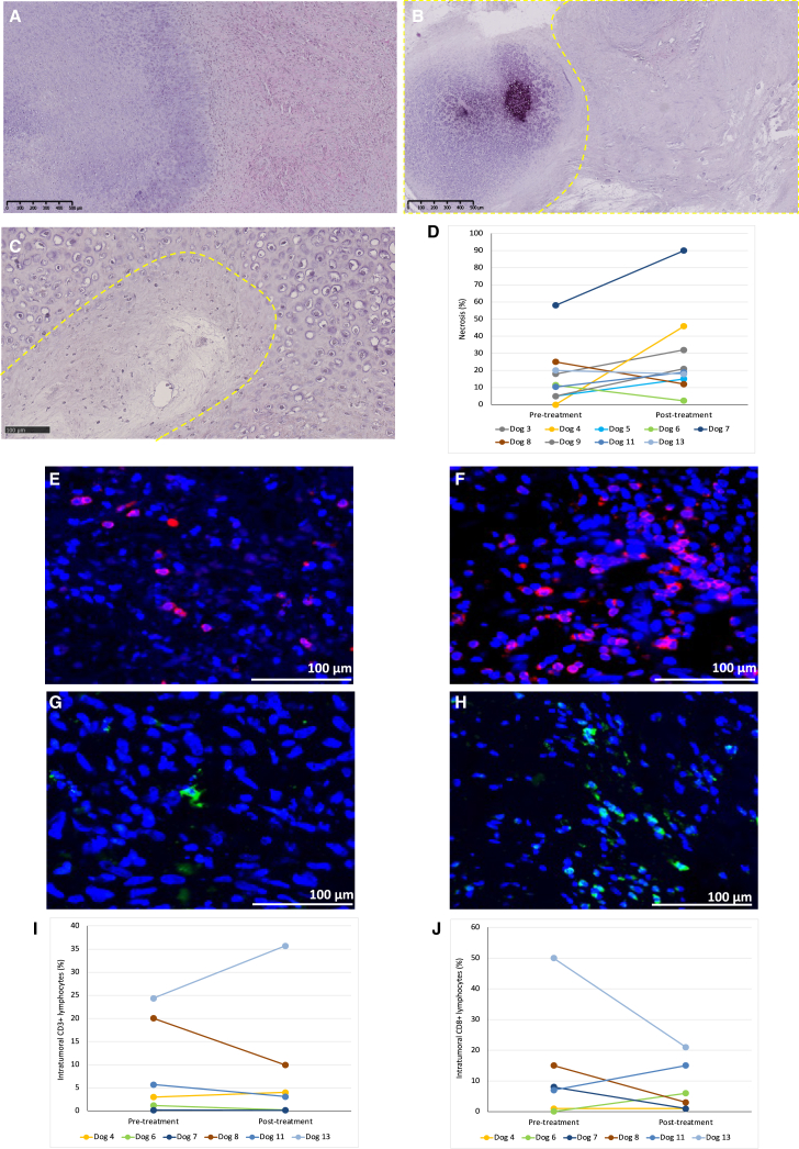
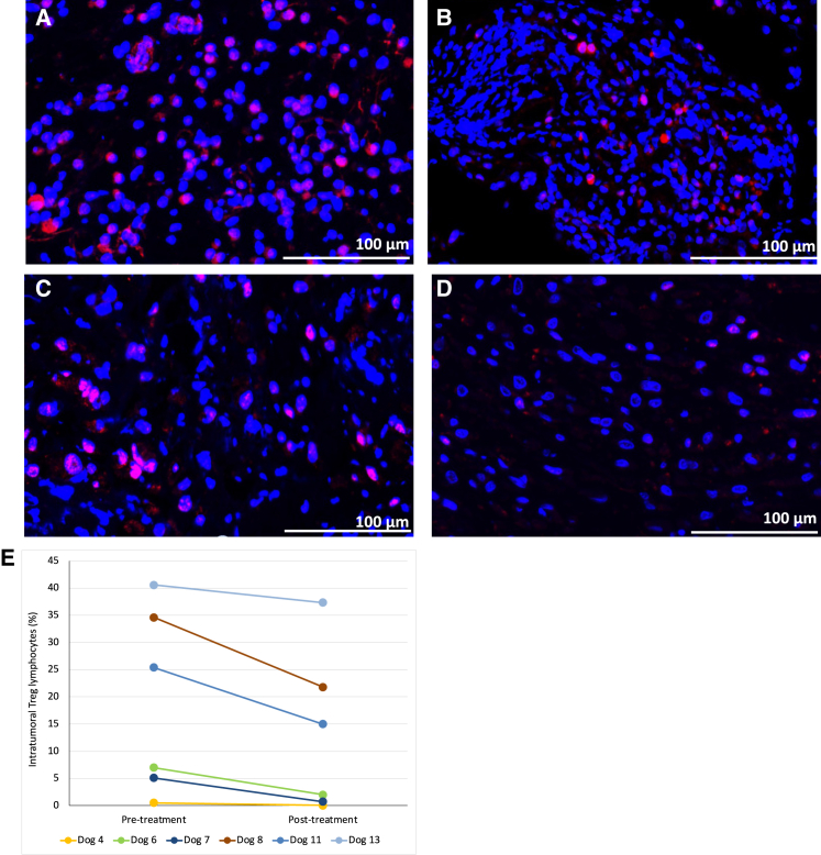
TG6002 is an oncolytic vaccinia virus expressing FCU1 protein, which converts 5-fluorocytosine into 5-fluorouracil. The study objectives were to assess tolerance, viral replication, 5-fluorouracil synthesis, and tumor microenvironment modifications to treatment in dogs with spontaneous malignant tumors. Thirteen dogs received one to three weekly intratumoral injections of TG6002 and 5-fluorocytosine. The viral genome was assessed in blood and tumor biopsies by qPCR. 5-Fluorouracil concentrations were measured in serum and tumor biopsies by liquid chromatography or high-resolution mass spectrometry. Histological and immunohistochemical analyses were performed. The viral genome was detected in blood (7/13) and tumor biopsies (4/11). Viral replication was suspected in 6/13 dogs. The median intratumoral concentration of 5-fluorouracil was 314 pg/mg. 5-Fluorouracil was not detected in the blood. An increase in necrosis (6/9) and a downregulation of intratumoral regulatory T lymphocytes (6/6) were observed. Viral replication, 5-fluorouracil synthesis, and tumor microenvironment changes were more frequently observed with higher TG6002 doses. This study confirmed the replicative properties, targeted chemotherapy synthesis, and reversion of the immunosuppressive tumor microenvironment in dogs with spontaneous malignant tumors treated with TG6002 and 5-fluorocytosine.
Keywords: antineoplastic protocols; immunomodulation; oncolytic virotherapy; oncolytic viruses; translational medical research.
© 2023 The Author(s).
Conflict of interest statement
J.B., S.C., M.G., I.F., C.P., B.M., J.F., J.-M.B., E.Q., and P.E. were employees of Transgene SA when the work was performed. Transgene SA is a publicly traded French biopharmaceutical company, with Institut Mérieux as the major shareholder. The authors declare no other competing interests. J.B. is a recipient of an Industrial Training Convention for Research (CIFRE) doctoral fellowship (2017/0266).
Figures


Similar articles
-
Safety studies and viral shedding of intramuscular administration of oncolytic vaccinia virus TG6002 in healthy beagle dogs.BMC Vet Res. 2020 Aug 25;16(1):307. doi: 10.1186/s12917-020-02524-y. BMC Vet Res. 2020. PMID: 32843040 Free PMC article.
-
Preclinical Evaluation of the Oncolytic Vaccinia Virus TG6002 by Translational Research on Canine Breast Cancer.Mol Ther Oncolytics. 2020 Sep 2;19:57-66. doi: 10.1016/j.omto.2020.08.020. eCollection 2020 Dec 16. Mol Ther Oncolytics. 2020. PMID: 33072863 Free PMC article.
-
The Enhanced Tumor Specificity of TG6002, an Armed Oncolytic Vaccinia Virus Deleted in Two Genes Involved in Nucleotide Metabolism.Mol Ther Oncolytics. 2019 Mar 27;14:1-14. doi: 10.1016/j.omto.2019.03.005. eCollection 2019 Sep 27. Mol Ther Oncolytics. 2019. PMID: 31011628 Free PMC article.
-
[Oncolytic virotherapy using replication-competent herpes simplex viruses].Uirusu. 2007 Jun;57(1):57-65. doi: 10.2222/jsv.57.57. Uirusu. 2007. PMID: 18040155 Review. Japanese.
-
Multidirectional Strategies for Targeted Delivery of Oncolytic Viruses by Tumor Infiltrating Immune Cells.Pharmacol Res. 2020 Nov;161:105094. doi: 10.1016/j.phrs.2020.105094. Epub 2020 Aug 12. Pharmacol Res. 2020. PMID: 32795509 Review.
Cited by
-
A Phase I Clinical Trial of Intrahepatic Artery Delivery of TG6002 in Combination with Oral 5-Fluorocytosine in Patients with Liver-Dominant Metastatic Colorectal Cancer.Clin Cancer Res. 2025 Apr 1;31(7):1243-1256. doi: 10.1158/1078-0432.CCR-24-2498. Clin Cancer Res. 2025. PMID: 39785814 Free PMC article. Clinical Trial.
-
A Review on Canine and Human Soft Tissue Sarcomas: New Insights on Prognosis Factors and Treatment Measures.Vet Sci. 2024 Aug 10;11(8):362. doi: 10.3390/vetsci11080362. Vet Sci. 2024. PMID: 39195816 Free PMC article. Review.
-
Combination of Oncolytic Virotherapy with Different Antitumor Approaches against Glioblastoma.Int J Mol Sci. 2024 Feb 7;25(4):2042. doi: 10.3390/ijms25042042. Int J Mol Sci. 2024. PMID: 38396720 Free PMC article. Review.
-
Oncolytic potential of Newcastle Disease Virus in feline lymphoma cells: an in vitro evaluation.Front Vet Sci. 2025 Jun 11;12:1484947. doi: 10.3389/fvets.2025.1484947. eCollection 2025. Front Vet Sci. 2025. PMID: 40567544 Free PMC article.
References
-
- Andtbacka R.H.I., Kaufman H.L., Collichio F., Amatruda T., Senzer N., Chesney J., Delman K.A., Spitler L.E., Puzanov I., Agarwala S.S., et al. Talimogene Laherparepvec Improves Durable Response Rate in Patients With Advanced Melanoma. J. Clin. Oncol. 2015;33:2780–2788. doi: 10.1200/JCO.2014.58.3377. - DOI - PubMed
-
- Foloppe J., Kempf J., Futin N., Kintz J., Cordier P., Pichon C., Findeli A., Vorburger F., Quemeneur E., Erbs P. The Enhanced Tumor Specificity of TG6002, an Armed Oncolytic Vaccinia Virus Deleted in Two Genes Involved in Nucleotide Metabolism. Mol. Ther. Oncolytics. 2019;14:1–14. doi: 10.1016/j.omto.2019.03.005. - DOI - PMC - PubMed
-
- Erbs P., Regulier E., Kintz J., Leroy P., Poitevin Y., Exinger F., Jund R., Mehtali M. In vivo cancer gene therapy by adenovirus-mediated transfer of a bifunctional yeast cytosine deaminase/uracil phosphoribosyltransferase fusion gene. Cancer Res. 2000;60:3813–3822. - PubMed
-
- Béguin J., Foloppe J., Maurey C., Laloy E., Hortelano J., Nourtier V., Pichon C., Cochin S., Cordier P., Huet H., et al. Preclinical evaluation of the oncolytic vaccinia virus TG6002 by translational research on canine breast cancer. Mol. Ther. Oncolytics. 2020;19:57–66. doi: 10.1016/j.omto.2020.08.020. - DOI - PMC - PubMed
LinkOut - more resources
Full Text Sources
