. 2023 Aug 12:6:100149.
doi: 10.1016/j.bioflm.2023.100149. eCollection 2023 Dec 15.

Octanoic acid promotes clearance of antibiotic-tolerant cells and eradicates biofilms of Staphylococcus aureus isolated from recurrent bovine mastitis

Affiliations

Octanoic acid promotes clearance of antibiotic-tolerant cells and eradicates biofilms of Staphylococcus aureus isolated from recurrent bovine mastitis

Wen-Chun Lin et al. Biofilm. .

Abstract

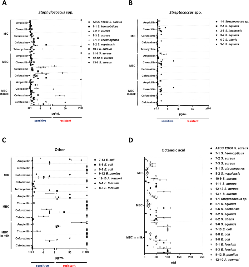
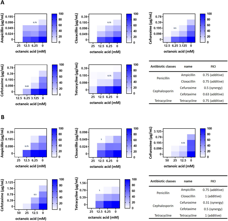
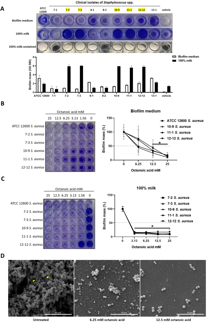
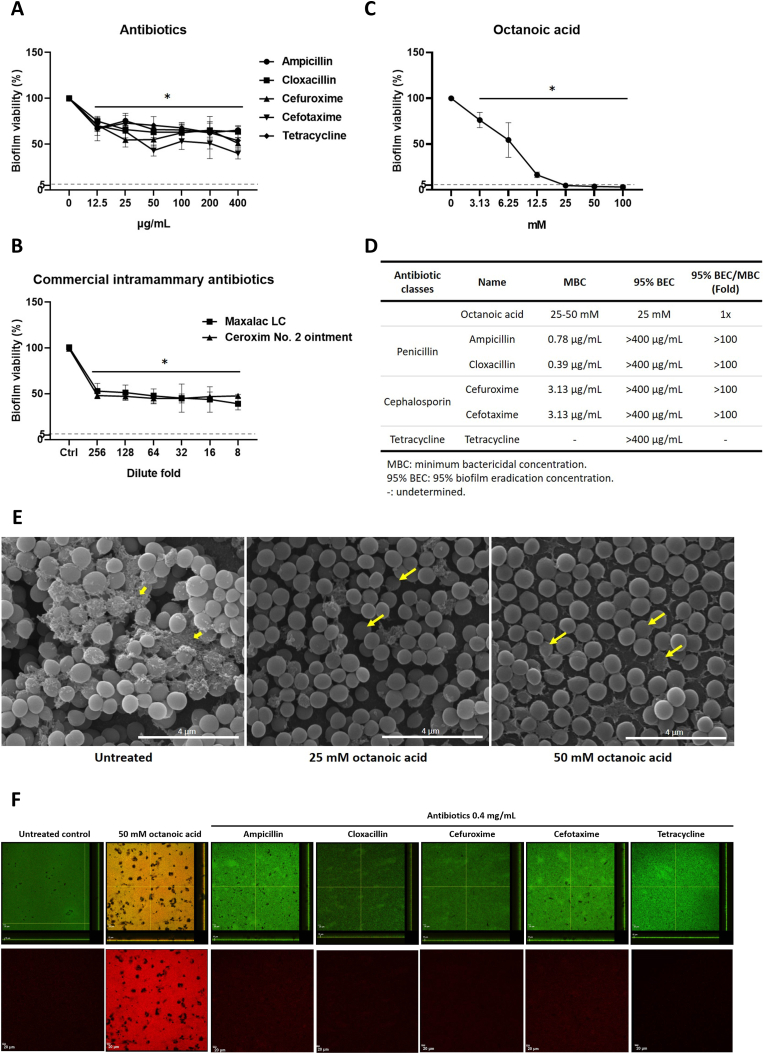
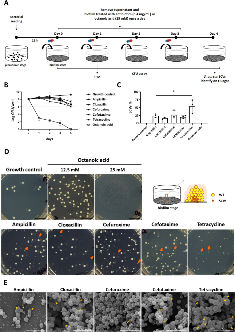
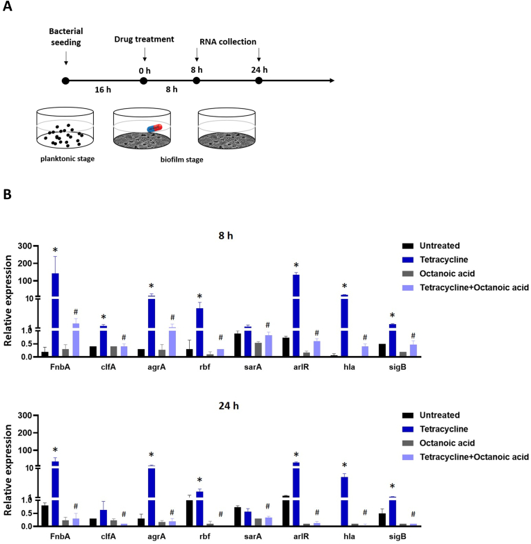
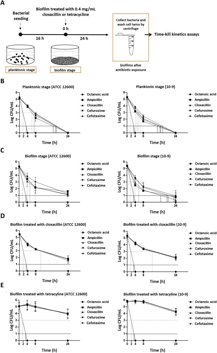
Antibiotic therapy is the primary treatment for bovine mastitis, but the drawbacks of this strategy include poor cure rate and economic losses from the need to discard milk with antibiotic residues. Unfortunately, few other treatment options are currently available for mastitis. Failure of antibiotic treatments is often attributed to formation of bacterial biofilms and abscesses in the mammary gland tissue, which lead to chronic infections that are difficult to eradicate and drive recurrent disease. A major mastitis-causing pathogen (MCP) associated with biofilms in bovine mastitis is Staphylococcus aureus. In this study, we demonstrate that octanoic acid has broad-spectrum microbicidal activity against MCPs and effectively inhibits S. aureus biofilm formation in milk (>50% inhibition at 3.13 mM). Octanoic acid effectively clears biofilms (95% eradication at 1X minimum bactericidal concentration, MBC) and infrequently induces S. aureus small colony variants (SCVs) that may cause recurrent mastitis. Additionally, octanoic acid rapidly kills persistent biofilm cells and cells with antibiotic tolerance (within 4 h). In contrast, antibiotics treated at >100X MBC cannot eradicate biofilms but do induce SCVs and antibiotic-tolerant cells. These effects may accelerate the transition from biofilm to chronic infection. Thus, octanoic acid exhibits bactericidal action against S. aureus biofilms, and it is less likely than antibiotic therapy to induce persistent cells and pathogen tolerance. Moreover, octanoic acid acts additively with antibiotics against S. aureus, and it attenuates tetracycline-induced virulence factor gene expression in S. aureus cells. According to these data, octanoic acid may prevent the pathological progression of bovine mastitis and offer a new strategy for treating the condition.

Keywords: Antibiotic tolerance cells; Octanoic acid; Persistence cells; Recurrent mastitis; Staphylococcus aureus biofilms.


Conflict of interest statement

The authors declare no competing interests.

Figures

Fig. 1
Fig. 1
Antibacterial activity of octanoic acid and antibiotics tested on bovine mastitis pathogens. Antibiotic susceptibility testing in (A) Staphylococcus spp., (B) Streptococcus spp., (C) Escherichia coli, and other pathogens isolated from dairy cows with mastitis. (D) Octanoic acid antimicrobial efficacy against S. aureus ATCC 12600 and clinical isolates of MCPs. MICs and MBCs of octanoic acid and antibiotics tested on S. aureus ATCC 12600 and clinical isolates of MCPs. The MBCs in milk were also determined in this study. All values represent the mean ± SD of three individual experiments. Determination of susceptibility of strains to antibiotics followed CLSI guidelines, with minor modifications. Susceptibility is defined by a MIC of ≤4 μg/mL and a MIC of ≥16 μg/mL was considered resistant.
Fig. 2
Fig. 2
Antibacterial effects of octanoic acid and antibiotic combinations against bovine mastitis pathogen S. aureus. Checkerboard analyses showing percentage inhibition for the combined effects of octanoic acid with ampicillin, cloxacillin, cefuroxime, cefotaxime and tetracycline toward (A) S. aureus ATCC 12600 and (B) clinical isolate 10-9. The heat map shows the average of three replicates. The positions of the blue values represent the concentrations calculated by FICI. (F) The FICI results and definitions of octanoic acid and antibiotic combinations. FICI ≤ 0.5 indicates synergism, while FICI >0.5–1 indicates an additive effect. (For interpretation of the references to colour in this figure legend, the reader is referred to the Web version of this article.)
Fig. 3
Fig. 3
Evaluation of biofilm formation inhibition activity of octanoic acid in the recurrence of mastitis major pathogens S. aureus. (A) A photograph of S. aureus ATCC 12600 and clinical isolates of Staphylococcus sp. biofilms stained with crystal violet (upper) and biofilm biomass quantification (lower). Highlighted (yellow) clinical isolates 7-2, 7-3, 10-9, 11-1, and 12-12 are strains with strong biofilm-forming capacity in milk. Biofilms formed by S. aureus ATCC 12600 and clinical isolates 7-2, 7-3, 10-9, 11-1, and 12-12 were tested with octanoic acid (0–25 mM) in sBHI (B) and milk (C). After a 24 h incubation at 37 °C, biofilms were stained with crystal violet for quantification of bacterial biomass. Untreated control was taken as 100% biofilm formation. The dotted line represents 50% biofilm inhibition. All values represent the mean ± SD of three individual experiments. *p < 0.05 compared with the untreated. (D) SEM images at 12000 × magnification in S. aureus ATCC 12600 biofilm formation treatment by octanoic acid (untreated, 6.25 and 12.5 mM). Yellow arrows represent the extracellular matrix of the S. aureus biofilm. Scale bars = 10 μm. (For interpretation of the references to colour in this figure legend, the reader is referred to the Web version of this article.)
Fig. 4
Fig. 4
Evaluation of octanoic acid and antibiotics in S. aureus biofilm eradication assay. (A) S. aureus ATCC 12600 mature biofilm treated with antibiotics (0–400 μg/mL), (B) commercial intramammary antibiotics (diluted 1:8 to 1:256 in sBHI), and (C) octanoic acid (0–100 mM) for 24 h. Biofilm cell viability was quantified by Alamar Blue assay. Untreated control was taken as 100% cell viability. The dotted line represents 95% biofilm eradication. Octanoic acid and antibiotic 95% biofilm eradication concentration (95% BEC) and 95% BEC/MBC are listed in table (D). All values represent the mean ± SD of three individual experiments. *p < 0.05 compared with the untreated control. (E) SEM images at 30,000 × magnification in ATCC 12600 S. aureus mature biofilm treatment by octanoic acid (untreated, 25 and 50 mM). Yellow arrows represent the ECM of S. aureus mature biofilm. Yellow thin arrows represent collapsed cells in octanoic acid-treated biofilms. Scale bars = 4 μm. (F) ATCC 12600 S. aureus mature biofilm cell viabilities were analyzed after culture in sBHI (Untreated), 50 mM octanoic acid, or 0.4 mg/mL antibiotics by CLSM at 20 × magnification. BacLight LIVE/DEAD viability kit was used to stain biofilm cells. Bacteria with intact cell membranes were stained fluorescent green (SYTO™ 9), while bacteria with damaged membranes were stained fluorescent red (propidium iodide). CLSM imaging shows green and red fluorescence (upper), representing the total cell content in biofilms, and red fluorescence alone (lower), representing the content of dead cells in biofilms. Lateral white scale bars = 20 μm. Longitudinal white scale bar = 15 μm. (For interpretation of the references to colour in this figure legend, the reader is referred to the Web version of this article.)
Fig. 5
Fig. 5
Antibiotic treatment induces SCV development in biofilms formed by S. aureus isolated from recurrent bovine mastitis. (A) Schematic diagram of the experimental process. Day 0 marks the day of mature biofilm formation. Mature biofilms of S. aureus isolate 10-9 were treated with 25 mM octanoic acid or 0.4 mg/mL antibiotic. Fresh drugs and sBHI (biofilm medium) were changed once a day until the third day. (B) Daily measurements of biofilm cell viability by CFU assay. The dotted line represents a decrease in cell viability by 2 log CFU compared to day 0. (C) Quantification of SCVs ratios (SCVs among total live cells) in different treatment groups (less than 1% is not detectable). SCVs were identified by LB agar plate method on the fourth day after drug treatment; SCVs were defined as colonies at least 10-fold smaller than normal colonies (growth control) [23]. All values represent the mean ± SD of three individual experiments. *p < 0.05 compared with the growth control. (D) The images of colony morphotypes on LB agar for each group (day 4). Scale bars = 1 cm. The orange arrows indicate SCV phenotypes developed upon exposure to antibiotics. (E) SEM analysis of morphological changes in biofilm cells after antibiotic treatment (day 1). The yellow arrows indicate SCV ‘fried egg' phenotypes [23] in the antibiotic treatment groups (compared with untreated control biofilm, see Fig. 4B). Images are magnified 30,000 × ; scale bars = 4 μm. (For interpretation of the references to colour in this figure legend, the reader is referred to the Web version of this article.)
Fig. 6
Fig. 6
Comparative transcript levels of virulence genes in S. aureus biofilms after treatment with octanoic acid, tetracycline, and octanoic acid/tetracycline combination. (A) Schematic diagram of the experimental procedure. (B) Mature biofilms of S. aureus isolate 10-9 were treated with 4 mM octanoic acid, 0.4 mg/mL tetracycline or a combination of both. After 8 h and 24 h incubation, biofilm cells were harvested for RNA extraction. Quantification of virulence gene (fnbA, clfA, agrA, rbf, sarA, arlR, hla and sigB) transcription in different treatment groups. Data are expressed as fold-change compared to 0 h (untreated). All values represent the mean ± SD of three individual experiments. *p < 0.05 compared with the untreated group (8 h and 24 h). #p < 0.05 compared with the tetracycline group (8 h and 24 h).
Fig. 7
Fig. 7
Comparative time-kill curves of octanoic acid and antibiotics against S. aureus cells at planktonic stage, biofilm stage, or biofilms after antibiotic (tetracycline or cloxacillin) exposure. (A) Schematic diagram of the experimental design. Representative time-kill curves for S. aureus ATCC 12600 and isolate 10-9 after exposure to octanoic acid at 2 × MBC (100 mM) and antibiotics at 4 × MBC (ampicillin, cloxacillin, cefuroxime and cefotaxime were respectively treated at 3.125, 1.56, 12.5, and 12.5 μg/mL for S. aureus ATCC 12600 and 1.56, 0.39, 3.13, and 6.25 μg/mL for S. aureus 10-9). S. aureus cells collected from the planktonic stage (B), biofilm stage (C), or biofilms after tetracycline (D) or cloxacillin (E) exposure. CFU/mL of each group was calculated at 0, 4, 8, and 24 h. The dotted line represents the minimum duration to kill 99% of the population (MDK99). The solid line represents the minimum duration to kill 99.99% of the population (MDK99.99). All values represent the mean ± SD of three individual experiments.

Similar articles

Cited by

References

    1. Gomes F., Henriques M. Control of bovine mastitis: old and recent therapeutic approaches. Curr Microbiol. 2016;72(4):377–382. doi: 10.1007/s00284-015-0958-8. - DOI - PubMed
    1. Tenhagen B.A., Köster G., Wallmann J., Heuwieser W. Prevalence of mastitis pathogens and their resistance against antimicrobial agents in dairy cows in Brandenburg, Germany. J Dairy Sci. 2006;89(7):2542–2551. doi: 10.3168/jds.S0022-0302(06)72330-X. - DOI - PubMed
    1. Krömker V., Leimbach S. Mastitis treatment-Reduction in antibiotic usage in dairy cows. Reprod Domest Anim. 2017;52(Suppl 3):21–29. doi: 10.1111/rda.13032. - DOI - PubMed
    1. Fisher R.A., Gollan B., Helaine S. Persistent bacterial infections and persister cells. Nat Rev Microbiol. 2017;15(8):453–464. doi: 10.1038/nrmicro.2017.42. - DOI - PubMed
    1. Peyrusson F., Varet H., Nguyen T.K., et al. Intracellular Staphylococcus aureus persisters upon antibiotic exposure. Nat Commun. 2020;11(1):2200. doi: 10.1038/s41467-020-15966-7. Published 2020 May 4. - DOI - PMC - PubMed

LinkOut - more resources