. 2023 Jul 27;26(9):107480.
doi: 10.1016/j.isci.2023.107480. eCollection 2023 Sep 15.

A tetracationic porphyrin with dual anti-prion activity

Affiliations

A tetracationic porphyrin with dual anti-prion activity

Antonio Masone et al. iScience. .

Erratum in

  • Erratum: A tetracationic porphyrin with dual anti-prion activity.
    Masone A, Zucchelli C, Caruso E, Lavigna G, Eraña H, Giachin G, Tapella L, Comerio L, Restelli E, Raimondi I, Elezgarai SR, De Leo F, Quilici G, Taiarol L, Oldrati M, Lorenzo NL, García-Martínez S, Cagnotto A, Lucchetti J, Gobbi M, Vanni I, Nonno R, Di Bari MA, Tully MD, Cecatiello V, Ciossani G, Pasqualato S, Van Anken E, Salmona M, Castilla J, Requena JR, Banfi S, Musco G, Chiesa R. Masone A, et al. iScience. 2023 Oct 18;26(11):108223. doi: 10.1016/j.isci.2023.108223. eCollection 2023 Nov 17. iScience. 2023. PMID: 37915608 Free PMC article.

Abstract

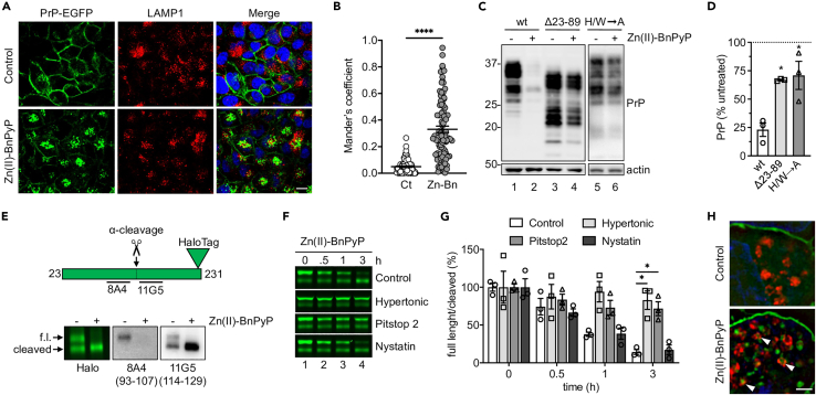
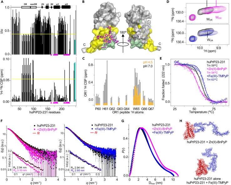
Prions are deadly infectious agents made of PrPSc, a misfolded variant of the cellular prion protein (PrPC) which self-propagates by inducing misfolding of native PrPC. PrPSc can adopt different pathogenic conformations (prion strains), which can be resistant to potential drugs, or acquire drug resistance, hampering the development of effective therapies. We identified Zn(II)-BnPyP, a tetracationic porphyrin that binds to distinct domains of native PrPC, eliciting a dual anti-prion effect. Zn(II)-BnPyP binding to a C-terminal pocket destabilizes the native PrPC fold, hindering conversion to PrPSc; Zn(II)-BnPyP binding to the flexible N-terminal tail disrupts N- to C-terminal interactions, triggering PrPC endocytosis and lysosomal degradation, thus reducing the substrate for PrPSc generation. Zn(II)-BnPyP inhibits propagation of different prion strains in vitro, in neuronal cells and organotypic brain cultures. These results identify a PrPC-targeting compound with an unprecedented dual mechanism of action which might be exploited to achieve anti-prion effects without engendering drug resistance.

Keywords: Cell biology; Molecular neuroscience; Pharmacology.


Conflict of interest statement

J.C. and H.E., as part of the company ATLAS Molecular Pharma S.L., declare that they have no conflicts of interest, as the company had no role in study design or funding, nor will they, or their immediate family members, benefit financially from the findings reported. All the other authors declare no competing interests.

Figures

None
Graphical abstract
Figure 1
Figure 1
Zn(II)-BnPyP down-regulates PrPC (A) Chemical structure of Zn(II)-BnPyP. (B) Chemical structure of Fe(III)-TMPyP. (C) Primary hippocampal neurons were treated with 0–10 μM Zn(II)-BnPyP for 24h and analyzed by Western blot (WB) with anti-PrP and anti-actin antibodies. (D) PrP signals were analyzed by densitometry of blots like the one shown in (C), normalized on the levels of actin, and expressed as percentages of untreated controls; mean ± SEM of 4–5 replicates from 5 independent experiments. (E) Neurons were treated with 2.5 μM Zn(II)-BnPyP for the times indicated and analyzed by WB. (F) Actin-normalized PrP levels; mean ± SEM of four experiments like the one shown in (E). (G) Confocal immunofluorescence analysis of PrP (green) and MAP2 (red) of neurons treated with 2.5 μM Zn(II)-BnP or Fe(III)-TMPyP for 6h. Cells were reacted with Hoechst 33258 (blue) to stain the nuclei. Scale bar 50 μm. (H) Neurons were exposed to 2.5 μM Zn(II)-BnPyP or Fe(III)-TMPyP for 24h, and analyzed by WB with anti-PrP and anti-actin antibodies. (I) Neurons treated with 2.5 μM Zn(II)-BnPyP for 24h were analyzed by WB to detect PrP, amyloid precursor protein (APP), tau, α-synuclein (α-syn) and actin. (J) H4 cells were incubated without (−) or with (+) 20 μM chloroquine and 5 μM Zn(II)-BnPyP, and analyzed by WB after 6h. (K) Actin-normalized PrP levels in H4 cells treated as in (J), relative to untreated controls, set as 100%; mean ± SEM. F3,16 = 7.443, p = 0.0024 by one-way ANOVA; ∗p < 0.05 vs. untreated, §§p < 0.01 vs. CQ, #p < 0.05 vs. Zn(II)-BnPyP by Tukey’s post-hoc test; CQ, chloroquine.
Figure 2
Figure 2
Zn(II)-BnPyP induces clathrin-dependent PrPC endocytosis and lysosomal degradation (A) Immunofluorescence analysis of LAMP1 in PrP-EGFP-expressing HeLa cells treated with the vehicle (control) or 5 μM Zn(II)-BnPyP, with 0.1 μg/μL bafilomycin A1 for 30 min. Scale bar 10 μm. (B) Mander’s coefficient M1: PrP-EGFP fraction that co-localizes with LAMP1. ∗∗∗∗p < 0.0001 by Student’s t test. Zn-Bn: Zn(II)-BnPyP. (C) Western blot of PrP and actin in HEK293 cells expressing moPrP wild-type (wt), Δ23-89, or with histidine- and tryptophan-to-alanine substitutions in the OR region (H/W→A), after 24h incubation without (−) or with (+) 5 μM Zn(II)-BnPyP. (D) Actin-normalized PrP levels in HEK293 cells relative to untreated controls, set as 100%. Mean ± SEM of three experiments like the one shown in C. ∗p < 0.05 by one-way ANOVA, Tukey’s post-hoc test. (E) Scheme of the PrP-Halo protein, indicating the site of α-cleavage and 8A4 and 11G5 antibody epitopes, and analysis of its expression in HEK293 cells visualized by cell impermeant Alexa Fluor 488 ligand fluorescence (Halo) or WB using 8A4 and 11G5 antibodies (epitopes in parentheses), before (−) and after (+) treatment with 5 μM Zn(II)-BnPyP for 6h. Arrows point to full-length (f.l.) and N-terminally cleaved PrP-Halo. (F) PrP-Halo visualized by cell impermeant Alexa Fluor 488 ligand in HEK293 cells treated with 5 μM Zn(II)-BnPyP for the times indicated, in standard (control) or hypertonic medium or with Pitstop 2 or nystatin. (G) Ratio of full-length to N-terminally cleaved PrP-Halo in cells treated as in (F). Mean ± SEM of three experiments. ∗p < 0.05 by two-way ANOVA, Dunnett’s post-hoc test. (H) SIM microscopy of PrP-Halo (green) and LAMP1 (red) in HEK293 cells before and after treatment with 5 μM Zn(II)-BnPyP for 6h with 0.1 μg/μL bafilomycin A1. Scale bar 2 μm.
Figure 3
Figure 3
Zn(II)-BnPyP inhibits replication of different prion strains in PMCA, N2a cells and COCS (A and B) Western blot analysis of PK-resistant PrP after a single 24h round of protein misfolding cyclic amplification (PMCA), with different dilutions of vCJD (A) or sCJD VV2 subtype (B) PrPSc seeds, with the vehicle or 5 μM Zn(II)-BnPyP or Fe(III)-TMPyP. Results are representative of two independent experiments. (C and D) WB analysis of total (-PK) and protease-resistant (+PK) PrP and actin in ScN2a-22L cells treated with 0, 0.5, 1, 2.5 and 5 μM Zn(II)-BnPyP or Fe(III)-TMPyP for 72h. (E) Quantification of actin-normalized PK-resistant PrP in ScN2a-22L cells treated with increasing concentrations of Zn(II)-BnPyP or Fe(III)-TMPyP. Data are expressed as percentages of untreated cells; mean ± SEM of three experiments. (F) WB of total (-PK) and protease-resistant (+PK) PrP in ScN2a-22L cells cultured without (−) or with (+) 5 μM Zn(II)-BnPyP for 16 days, then without porphyrin (wash-out) for the days indicated. (G) WB of PrP and actin in RML- and 22L-infected COCS treated with Zn(II)-BnPyP at the concentrations indicated. Blots were overexposed to show the presence of full-length PrPC and the absence of PK-resistant PrP in the treated COCS (shorter exposure of a replicate experiment is shown in Figure S4F).
Figure 4
Figure 4
Structural characterization of the Zn(II)-BnPyP interaction with huPrP23-231 (A) Intensity ratio (I/Io) and 1H15N chemical shift perturbations (CSPs) of 15N huPrP23-231 amide resonances on addition of Zn(II)-BnPyP (1:2). Magenta triangles: peaks that disappeared; yellow line: avg - sd (I/Io) or avg + sd (CSP); cyan bars: data refer to OR W indole NH. (B) Docking model of huPrP23-231 globular domain in complex with Zn(II)-BnPyP. Yellow: residues whose resonances are significantly affected by Zn(II)-BnPyP; pale green: unassigned resonances because of overlap or solvent exchange. (C) CSPs of OR1 peptide 1H resonances on addition of Zn(II)-BnPyP (1:1) at pH 7 (black) or pH 4.5 (orange). (D) Superposition of 1H-15N HSQC spectra of 15N huPrP23-231 without (black) and with (1:1) Zn(II)-BnPyP (magenta) or Fe(III)-TMPyP (blue), zoom into tryptophan (W) indole NH region. (E) Circular dichroism melting curves of huPrP23-231 alone (black), on addition (1:15) of Zn(II)-BnPyP (magenta) or Fe(III)-TMPyP (blue). Mean of two replicates ±SEM. (F) SAXS curves of huPrP23-231 alone (black) and in complex (1:5) with Zn(II)-BnPyP (magenta) or Fe(III)-TMPyP (blue); red: GNOM fitting curve. The insets show the Guinier fits. (G) Pair distance distributions of huPrP23-231 without (black) and with (1:5) Zn(II)-BnPyP (magenta) or Fe(III)-TMPyP (blue). (H) Illustrative protein structures after EOM modeling of SAXS data, describing huPrP23-231 in the presence of Zn(II)-BnPyP (upper panel) and alone or with Fe(III)-TMPyP (lower panel). The C-terminal globular domain (red) was used as a rigid body and the N-terminal tail (blue) was modeled by EOM.
Figure 5
Figure 5
Zn(II)-BnPyP pharmacokinetics and effect on brain PrPC levels (A and B) C57BL/6 mice were injected ip with 10 mg/kg Zn(II)-BnPyP and euthanized at different times after the dose (5 min, 30 min, 1h, 2h, 6h, 24h, 3 days and 7 days). Porphyrin in the plasma (A) and the brain (B) was quantified by HPLC coupled with a fluorescence detector. Data are the mean ± SD of three mice. (C) C57BL/6 mice were injected ip with 10 mg/kg Zn(II)-BnPyP every second day for the weeks indicated, and euthanized 6h after the last dose for quantification of brain Zn(II)-BnPyP. Data are the mean ± SD of 3–5 mice. (D–F) Quantification (mean ± SD) of actin-normalized PrPC levels in the brains of the mice treated for 4, 8 or 12 weeks shown in C.
Figure 6
Figure 6
Zn(II)-BnPyP does not affect RML-induced prion disease (A and B) Kaplan-Meier plots showing time to onset (A) and to terminal disease (B) of C57BL/6 mice intracerebrally inoculated with RML and treated ip with saline (vehicle; n = 6) or 10 mg/kg Zn(II)-BnPyP (n = 9) every second day from 40 dpi. (C) Representative WB analysis of PK-resistant PrP in the brains of RML-inoculated mice, treated with vehicle or Zn(II)-BnPyP and euthanized at the terminal stage of the disease. Brain extracts were digested with 0, 5, 25, and 50 μg/mL of PK and analyzed by WB with anti-PrP 12B2 monoclonal antibody. The undigested samples (0 μg/mL PK) amount to 10 μg of protein, and the other samples 20 μg.

References

    1. Colby D.W., Prusiner S.B. Prions. Cold Spring Harb. Perspect. Biol. 2011;3:a006833. doi: 10.1101/cshperspect.a006833. - DOI - PMC - PubMed
    1. Chiesa R. The elusive role of the prion protein and the mechanism of toxicity in prion disease. PLoS Pathog. 2015;11:e1004745. doi: 10.1371/journal.ppat.1004745. - DOI - PMC - PubMed
    1. Harris D.A. Trafficking, turnover and membrane topology of PrP. Br. Med. Bull. 2003;66:71–85. - PubMed
    1. Zahn R., Liu A., Lührs T., Riek R., von Schroetter C., López García F., Billeter M., Calzolai L., Wider G., Wüthrich K. NMR solution structure of the human prion protein. Proc. Natl. Acad. Sci. USA. 2000;97:145–150. doi: 10.1073/pnas.97.1.145. - DOI - PMC - PubMed
    1. Spevacek A.R., Evans E.G.B., Miller J.L., Meyer H.C., Pelton J.G., Millhauser G.L. Zinc drives a tertiary fold in the prion protein with familial disease mutation sites at the interface. Structure. 2013;21:236–246. doi: 10.1016/j.str.2012.12.002. - DOI - PMC - PubMed