Review
. 2023 Aug 10:10:1232882.
doi: 10.3389/fcvm.2023.1232882. eCollection 2023.

Clinical magnetocardiography: the unshielded bet-past, present, and future

Affiliations
Review

Clinical magnetocardiography: the unshielded bet-past, present, and future

D Brisinda et al. Front Cardiovasc Med. .

Abstract

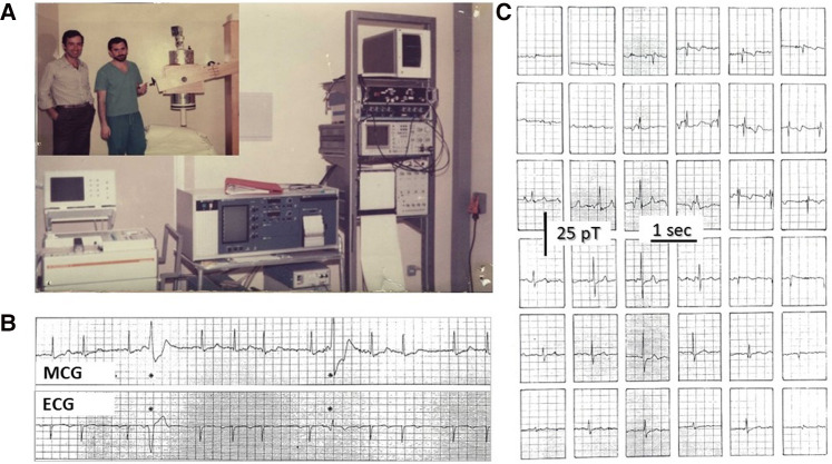
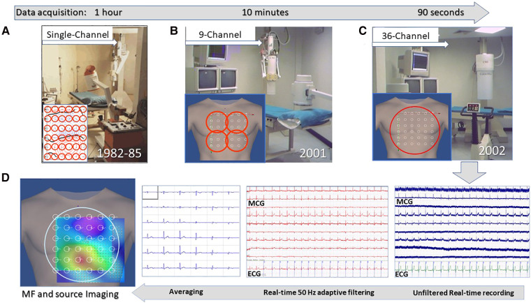
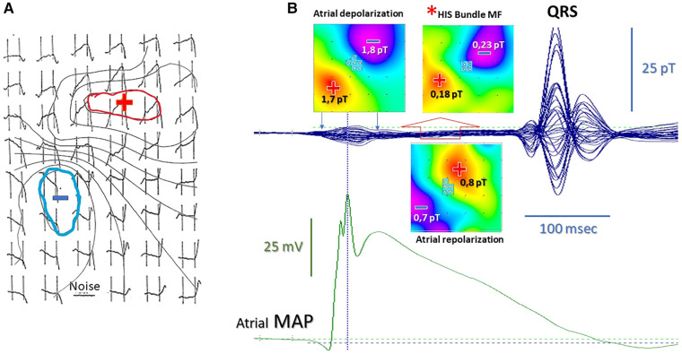
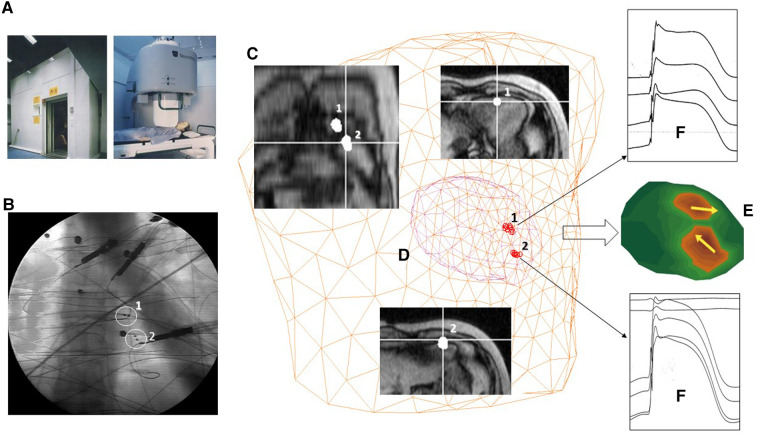
Magnetocardiography (MCG), which is nowadays 60 years old, has not yet been fully accepted as a clinical tool. Nevertheless, a large body of research and several clinical trials have demonstrated its reliability in providing additional diagnostic electrophysiological information if compared with conventional non-invasive electrocardiographic methods. Since the beginning, one major objective difficulty has been the need to clean the weak cardiac magnetic signals from the much higher environmental noise, especially that of urban and hospital environments. The obvious solution to record the magnetocardiogram in highly performant magnetically shielded rooms has provided the ideal setup for decades of research demonstrating the diagnostic potential of this technology. However, only a few clinical institutions have had the resources to install and run routinely such highly expensive and technically demanding systems. Therefore, increasing attempts have been made to develop cheaper alternatives to improve the magnetic signal-to-noise ratio allowing MCG in unshielded hospital environments. In this article, the most relevant milestones in the MCG's journey are reviewed, addressing the possible reasons beyond the currently long-lasting difficulty to reach a clinical breakthrough and leveraging the authors' personal experience since the early 1980s attempting to finally bring MCG to the patient's bedside for many years thus far. Their nearly four decades of foundational experimental and clinical research between shielded and unshielded solutions are summarized and referenced, following the original vision that MCG had to be intended as an unrivaled method for contactless assessment of the cardiac electrophysiology and as an advanced method for non-invasive electroanatomical imaging, through multimodal integration with other non-fluoroscopic imaging techniques. Whereas all the above accounts for the past, with the available innovative sensors and more affordable active shielding technologies, the present demonstrates that several novel systems have been developed and tested in multicenter clinical trials adopting both shielded and unshielded MCG built-in hospital environments. The future of MCG will mostly be dependent on the results from the ongoing progress in novel sensor technology, which is relatively soon foreseen to provide multiple alternatives for the construction of more compact, affordable, portable, and even wearable devices for unshielded MCG inside hospital environments and perhaps also for ambulatory patients.

Keywords: electroanatomical imaging; electrophysiology arrhythmias mapping and ablation; gradiometer array; inverse problem; magnetic sensors; magnetically shielded room (MSR); magnetocardiography (MCG); source localization accuracy.


Conflict of interest statement

The authors declare that the research was conducted in the absence of any commercial or financial relationships that could be construed as a potential conflict of interest.

Figures

Figure 1
Figure 1
(A) CNR's physicist Gian Luca Romani (left) and Riccardo Fenici (right) testing the prototype of the CNR unshielded MCG SQUID gradiometer at the Catholic University's Gemelli Hospital in Rome. (B) Example of real-time simultaneous MCG and ECG recordings and (C) example of 36-position real-time single-beat MCG recordings (Finnish grid) [modified from (98)].
Figure 2
Figure 2
Unshielded magnetocardiography at the Catholic University Hospital's BACPIC: (A) Single-channel system; (B) Nine-channel CMI prototype; (C) CMI 3619a 36-channel; and (D) examples of real-time MCG recordings and of the signal processing flow chart for MF imaging and 3D source localization.
Figure 3
Figure 3
(A) High-resolution MCG map of the “ramp-like” pattern during the PR interval with superimposed isomagnetic lines [modified from (120) data]. (B) Validation of the atrial repolarization nature of the PR “ramp-like” MCG pattern with simultaneous MCG and atrial monophasic action potential recording. The red asterisk indicates the His bundle MF (±0.2pT) disclosed during the last half of the PR interval after subtraction of the stronger (±1.8 pT) atrial repolarization MF (open red arrow). The pseudo-current reconstruction (multiple white arrows on the MF maps) indicates the opposite direction of atrial and His bundle depolarization current and of atrial repolarization current [standardized MF color code: in blue (−), MF outgoing from the chest; in red (+), MF entering in the chest].
Figure 4
Figure 4
Example of multimodal 3D EAI based on MSI validated in the Helsinki University Central Hospital's BioMag Laboratory. (A) The MSR. (B) Fluoroscopic imaging of two amagnetic catheters (ACs). (C) MRI imaging. (D) 3D localization of the distal end of the ACs with the MCG ECD inverse solution. (E) 3D current density imaging of two 10 µA current dipoles generated by the ACs. (F) Multiple monophasic action potential recordings from the ACs.
Figure 5
Figure 5
Example of non-invasive MCG localization of a right posterior-septal Kent bundle during sustained antidromic reentry tachycardia induced by transesophageal atrial pacing and spontaneous desynchronization in atrial fibrillation. The accessory pathway localization was confirmed by successful RF ablation.
Figure 6
Figure 6
Typical experimental setup used for the MCG study of small animals with the CMI 3649a system. (A) Non-invasive contactless MCG recording and source imaging. (B) Procedure for simultaneous minimally invasive electrophysiological study with a single AC for multiple monophasic action potential recording from the epicardial surface, localizable with the MCG mapping and EMD inverse solution [red-circled solid arrow in (C)].
Figure 7
Figure 7
Examples of most recent novel MCG devices, two operating unshielded: (A) the cryogenic Avalon-H90. (B) The portable VitalScan/Corsens and one still requiring local EMS provided by a more compact cylindric MSR. (C) The CardioFlux, featuring zero-field OPMs.
Figure 8
Figure 8
First reported comparison between sequential 36-point MCG recording with a single-channel OPM developed at the University of Freiburg (Swiss) and 36-channel MCG mapping at the Catholic University Hospital in Rome. All data were converted to the same format and analyzed with the Neuromag MCG software. The identity of MCG waveforms, of the MF distribution, of the related source localization (green arrows onto the MF maps), and of the 3D current density imaging (CDI), calculated at the apex of the QRS and of the T waves, is immediately evident [modified from data in (275)].
Figure 9
Figure 9
First reported test of a dual-channel scalar field OPMs (geometrics MFAM) arranged as a first-order gradiometer (baseline 5 cm) in the Catholic University Hospital's unshielded BACPIC: (A) Preliminary phantom measurement of a 1 mA current dipole generated by the amagnetic catheter. (B) Comparison between MCG recording and CMI 36-channel MCG mapping (R-wave MF distribution). The similarity of MCG waveforms recorded with the MFAM (B1) and with the SQUID (B2) at the same grid position (white circle onto the MF map) is evident. The amplitude of the MCG signal recorded with the MFAM is higher because the OPMs can be placed closer to the chest. (C) Noise spectra of each MFAM and of the first-order gradiometer.

References

    1. Plonsey R. Comparative capabilities of electrocardiography and magnetocardiography. Am J Cardiol. (1972) 29(5):735–6. 10.1016/0002-9149(72)90179-8 - DOI - PubMed
    1. Malmivuo J, Plonsey R. Bioelectromagnetism—principles and applications of bioelectric and biomagnetic fields (1995). BioLabor Biofizikai és Laboratóriumi Szolg. Kft. Available at: www.biolabor.hu. (last accessed 02 July 2023)
    1. Grynszpan F, Geselowitz DB. Model studies of the magnetocardiogram. Biophys J. (1973) 13(9):911–25. 10.1016/S0006-3495(73)86034-5 - DOI - PMC - PubMed
    1. Wikswo JPJ, Barach JP. Possible sources of new information in the magnetocardiogram. J Theor Biol. (1982) 95(4):721–9. 10.1016/0022-5193(82)90350-2 - DOI - PubMed
    1. Roth BJ, Wikswo JP. Electrically silent magnetic fields. Biophys J. (1986) 50(4):739–45. 10.1016/S0006-3495(86)83513-5 - DOI - PMC - PubMed