

Finite element modeling of the human cervical spinal cord and its applications: A systematic review
- PMID: 37636342
- PMCID: PMC10448221
- DOI: 10.1016/j.xnsj.2023.100246
Finite element modeling of the human cervical spinal cord and its applications: A systematic review
Abstract
Background context: Finite element modeling (FEM) is an established tool to analyze the biomechanics of complex systems. Advances in computational techniques have led to the increasing use of spinal cord FEMs to study cervical spinal cord pathology. There is considerable variability in the creation of cervical spinal cord FEMs and to date there has been no systematic review of the technique. The aim of this study was to review the uses, techniques, limitations, and applications of FEMs of the human cervical spinal cord.
Methods: A literature search was performed through PubMed and Scopus using the words finite element analysis, spinal cord, and biomechanics. Studies were selected based on the following inclusion criteria: (1) use of human spinal cord modeling at the cervical level; (2) model the cervical spinal cord with or without the osteoligamentous spine; and (3) the study should describe an application of the spinal cord FEM.
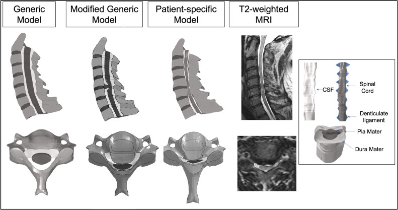
Results: Our search resulted in 369 total publications, 49 underwent reviews of the abstract and full text, and 23 were included in the study. Spinal cord FEMs are used to study spinal cord injury and trauma, pathologic processes, and spine surgery. Considerable variation exists in the derivation of spinal cord geometries, mathematical models, and material properties. Less than 50% of the FEMs incorporate the dura mater, cerebrospinal fluid, nerve roots, and denticulate ligaments. Von Mises stress, and strain of the spinal cord are the most common outputs studied. FEM offers the opportunity for dynamic simulation, but this has been used in only four studies.
Conclusions: Spinal cord FEM provides unique insight into the stress and strain of the cervical spinal cord in various pathological conditions and allows for the simulation of surgical procedures. Standardization of modeling parameters, anatomical structures and inclusion of patient-specific data are necessary to improve the clinical translation.
Keywords: Biomechanics; Cervical spinal cord; Finite element modeling; Ligamentum flavum hypertrophy; Ossification of the posterior longitudinal ligament; Spinal cord injury; Spine surgery; Trauma.
Conflict of interest statement
The authors declare that they have no known competing financial interests or personal relationships that could have appeared to influence the work reported in this paper
Figures


References
Publication types
LinkOut - more resources
Full Text Sources
