. 2023 Aug 11:17:1219847.
doi: 10.3389/fncel.2023.1219847. eCollection 2023.

Defective hippocampal neurogenesis underlies cognitive impairment by carotid stenosis-induced cerebral hypoperfusion in mice

Affiliations

Defective hippocampal neurogenesis underlies cognitive impairment by carotid stenosis-induced cerebral hypoperfusion in mice

Enrique Fraga et al. Front Cell Neurosci. .

Abstract

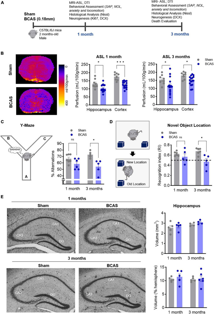
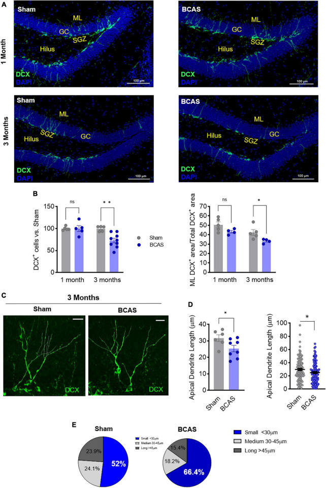
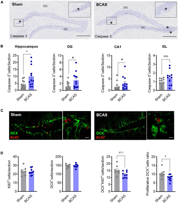
Chronic cerebral hypoperfusion due to carotid artery stenosis is a major cause of vascular cognitive impairment and dementia (VCID). Bilateral carotid artery stenosis (BCAS) in rodents is a well-established model of VCID where most studies have focused on white matter pathology and subsequent cognitive deficit. Therefore, our aim was to study the implication of adult hippocampal neurogenesis in hypoperfusion-induced VCID in mice, and its relationship with cognitive hippocampal deficits. Mice were subjected to BCAS; 1 and 3 months later, hippocampal memory and neurogenesis/cell death were assessed, respectively, by the novel object location (NOL) and spontaneous alternation performance (SAP) tests and by immunohistology. Hypoperfusion was assessed by arterial spin labeling-magnetic resonance imaging (ASL-MRI). Hypoperfused mice displayed spatial memory deficits with decreased NOL recognition index. Along with the cognitive deficit, a reduced number of newborn neurons and their aberrant morphology indicated a remarkable impairment of the hippocampal neurogenesis. Both increased cell death in the subgranular zone (SGZ) and reduced neuroblast proliferation rate may account for newborn neurons number reduction. Our data demonstrate quantitative and qualitative impairment of adult hippocampal neurogenesis disturbances associated with cerebral hypoperfusion-cognitive deficits in mice. These findings pave the way for novel diagnostic and therapeutic targets for VCID.

Keywords: aberrant neurogenesis; carotid stenosis; dementia; hippocampus; hypoperfusion; maladaptive remodeling; neurogenesis; vascular cognitive impairment.


Conflict of interest statement

The authors declare that the research was conducted in the absence of any commercial or financial relationships that could be construed as a potential conflict of interest.

Figures

FIGURE 1
FIGURE 1
Chronic cerebral hypoperfusion induces hippocampus-memory impairment in mice without signs of hippocampal atrophy. (A) Experimental design. (B) Arterial spin labeling (ASL) was used to measure CBF after 1 (middle) or 3 months (right) of BCAS in hippocampus and cortex. Representative pictures 3 months after surgery are shown (left) (n = 8, 16). (C) Percentage of spontaneous alternation in the Y maze at 1–3 months of BCAS (1 month, n = 6–7, 3 months, n = 5). (D) NOL test results expressed as recognition index (n = 5). (E) Volumetric assessment of hippocampus, normalized by hemisphere volume (n = 4–5). Representative Nissl-stained images. Data are mean ± SEM. Statistical assessment by ANOVA and multiple Mann-Whitney tests with Holm-Sidak correction for the paired comparisons. *p < 0.05, ***p < 0.001.
FIGURE 2
FIGURE 2
Memory deficits are concomitant to impaired hippocampal neurogenesis following hypoperfusion. (A) Representative images of doublecortin (DCX) staining in the DG. Scale bar = 100 μm. (B) DCX+ cells number (left) and % of DCX+ area in the molecular layer (ML; right) after BCAS (n = 4–9). (C) Representative high-resolution images of DCX+ cells. Scale bar = 15 μm. (D) Quantification of apical dendrite length. Left: mean apical dendrite by animal; right: distribution of every apical dendrite length (n = 6–8). (E) Pie charts of the distribution in apical dendrite length. Statistical assessment by ANOVA and multiple Mann-Whitney tests with Holm-Sidak correction for the paired comparisons. Two comparisons were tested by Mann-Whitney U-test. *p < 0.05, **p < 0.01.
FIGURE 3
FIGURE 3
Mechanisms leading to impaired hippocampal neurogenesis. (A) Representative images of cleaved caspase-3 in hippocampus 3 months after surgery. Scale bar = 250 and 50 μm. (B) Cell death quantification in different hippocampal regions. Mann-Whitney test was used to compare the ranks (n = 12). (C) Representative images of DCX (green) and Ki-67 (red) co-staining in DG 1 month after surgery. Scale bars = 80 and 15 μm (n = 7–9). (D) Quantification of Ki67+, DCX+, and Ki67+/DCX+ cells and of the proliferation ratio of DCX+ cells. Two-tailed Mann-Whitney U-test was used to compare the ranks. *p < 0.05.

References

    1. Alonso-Alconada D., Gressens P., Golay X., Robertson N. J. (2022). Neurogenesis is reduced at 48 h in the subventricular zone independent of cell death in a piglet model of perinatal hypoxia-ischemia. Front. Pediatr. 10:793189. 10.3389/fped.2022.793189 - DOI - PMC - PubMed
    1. Berry C., Sidik N., Pereira A. C., Ford T. J., Touyz R. M., Kaski J. C., et al. (2019). Small-vessel disease in the heart and brain: Current knowledge, unmet therapeutic need, and future directions. J. Am. Heart Assoc. 8:e011104. 10.1161/JAHA.118.011104 - DOI - PMC - PubMed
    1. Boehm-Sturm P., Füchtemeier M., Foddis M., Mueller S., Trueman R. C., Zille M., et al. (2017). Neuroimaging biomarkers predict brain structural connectivity change in a mouse model of vascular cognitive impairment. Stroke 48 468–475. 10.1161/STROKEAHA.116.014394 - DOI - PMC - PubMed
    1. Carter R. J., Lione L. A., Humby T., Mangiarini L., Mahal A., Bates G. P., et al. (1999). Characterization of progressive motor deficits in mice transgenic for the human Huntington’s disease mutation. J. Neurosci. 19, 3248–3257. 10.1523/JNEUROSCI.19-08-03248.1999 - DOI - PMC - PubMed
    1. Coltman R., Spain A., Tsenkina Y., Fowler J. H., Smith J., Scullion G., et al. (2011). Selective white matter pathology induces a specific impairment in spatial working memory. Neurobiol. Aging 32:2324.e7–12. 10.1016/j.neurobiolaging.2010.09.005 - DOI - PubMed