

Borderline Malignant Sebaceoma of the Auricle: A Case Report
- PMID: 37636626
- PMCID: PMC10447812
- DOI: 10.1007/s12070-023-03552-4
Borderline Malignant Sebaceoma of the Auricle: A Case Report
Abstract
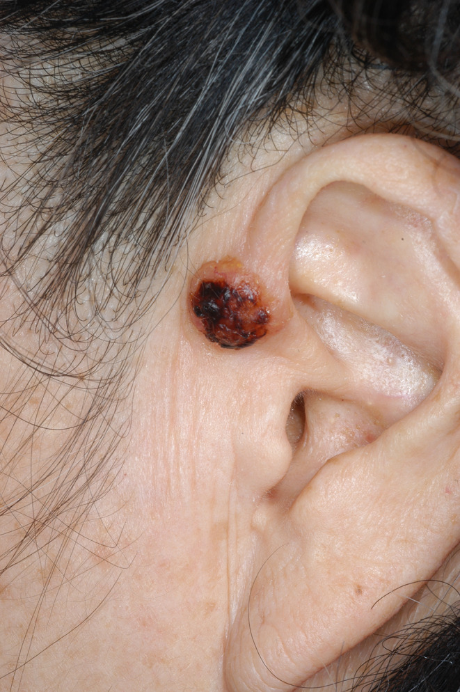
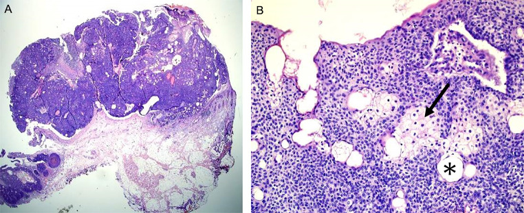
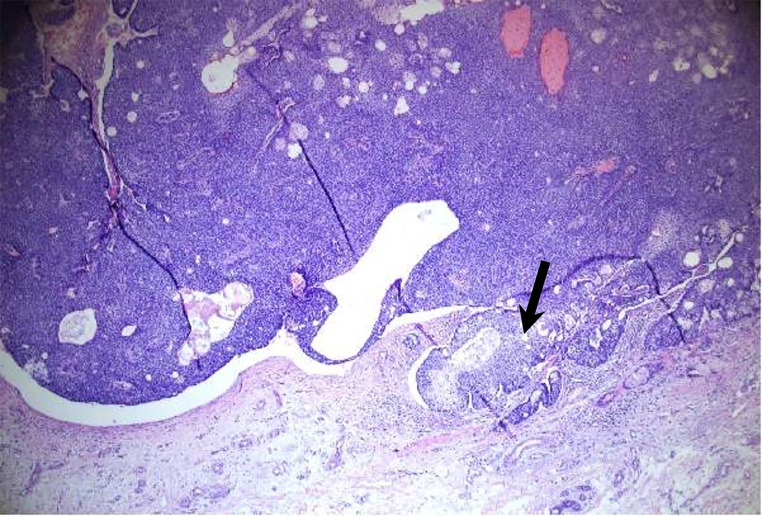
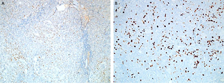
Sebaceoma is a rare benign tumor arising from the sebaceous gland of the skin. Sebaceoma often occurs on the head and neck but rarely on the ears. We present the case of a 78-year-old female patient with a two-year history of a protruding mass in her left ear. Physical examination revealed a well-circumscribed plaque in the crus of the helix of the left ear. A wide local excisional biopsy was taken, and the mass was subjected to histopathologic assessment. While the mass showed cytological findings indicating sebaceoma, it also presented malignant features architecturally and immunohistochemically. Based on these findings, the tumor was regarded as a sebaceoma of borderline malignancy.
Supplementary information: The online version contains supplementary material available at 10.1007/s12070-023-03552-4.
Keywords: Ear Auricle; Malignancy; Sebaceoma; Sebaceous Gland.
© Association of Otolaryngologists of India 2023. Springer Nature or its licensor (e.g. a society or other partner) holds exclusive rights to this article under a publishing agreement with the author(s) or other rightsholder(s); author self-archiving of the accepted manuscript version of this article is solely governed by the terms of such publishing agreement and applicable law.
Conflict of interest statement
Conflict of interestAuthors declare that they have no conflict of interest.
Figures
References
LinkOut - more resources
Full Text Sources