Small Round Blue Cell Tumours of the Sinonasal Area: Our 5 year Experience in a Tertiary Care Centre in India
- PMID: 37636680
- PMCID: PMC10447677
- DOI: 10.1007/s12070-023-03840-z
Small Round Blue Cell Tumours of the Sinonasal Area: Our 5 year Experience in a Tertiary Care Centre in India
Abstract
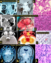
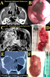
Purpose: The main purpose of this study is to understand the characteristics and management of sinonasal small round blue cell tumors and also to emphasise the role of immunohistochemistry in their diagnosis and on the outcomes after endoscopic/open excision in these patients. Methods: This is a retrospective study conducted at a tertiary care referral centre in India which included 38 patients with sino nasal for a period of 5 years. All the patients were evaluated clinically and radiologically. All cases were confirmed diagnostically with histopathological examination and immunohistochemistry following surgical excision either by endoscopic or open approach. Some of the cases underwent post operative radiotherapy. Results: In our study, among 176 cases diagnosed with Sino nasal malignancies, 38 (21.6%) cases were diagnosed with sinonasal small round blue cell tumors with male to female ratio 1.4:1. Most common histopathological type among all the sinonasal small round blue cell tumors that presented to us was esthesioneuroblastoma i.e., 8 (21%) patients followed by pituitary macroadenoma in 7(8.4%) patients. Other types are undifferentiated squamous cell carcinoma 10(13.1%), craniopharyngioma 8(10.5%), lymphoma 3(7.9%), synovial/spindle cell sarcoma, malignant melanoma and adenocarcinoma 1(2.6%) each. Schwannoma, rhabdomyosarcoma, neuroendocrine carcinoma and neurofibroma 2 (5.2%) each. Conclusion: Sinonasal small round blue cell tumors are extremely rare tumours. Histopathological diagnosis with immunohistochemistry is characteristic of various tumors and is conclusive for diagnosis. Knowledge of these tumor entity is essential as early diagnosis helps in further management in preventing spread to vital structures and improving outcome. Most of the tumors have a multimodality treatment approach which includes surgical excision, radiotherapy and chemotherapy.
Keywords: Endoscopic excision; Esthesioneuroblastoma; Malignant melanoma; Radiotherapy; Rhabdomyosarcoma; SRBCT’s immunohistochemistry; Schwannoma; Sinonasal malignancies; Sinonasal undifferentiated carcinoma; Small round blue cell tumours; Spindle cell tumour; Synovial sarcoma.
© Association of Otolaryngologists of India 2023. Springer Nature or its licensor (e.g. a society or other partner) holds exclusive rights to this article under a publishing agreement with the author(s) or other rightsholder(s); author self-archiving of the accepted manuscript version of this article is solely governed by the terms of such publishing agreement and applicable law.
Conflict of interest statement
Conflict of InterestThe author(s) declared no potential conflict of interest with respect to the research, authorship, and/or publication of this article.
Figures


Similar articles
-
The Small Round Blue Cell Tumors of Sinonasal Tract: Pathologists Grey Zone.J Microsc Ultrastruct. 2022 Feb 4;12(1):21-26. doi: 10.4103/jmau.jmau_70_21. eCollection 2024 Jan-Mar. J Microsc Ultrastruct. 2022. PMID: 38633570 Free PMC article.
-
"Undifferentiated" small round cell tumors of the sinonasal tract: differential diagnosis update.Am J Clin Pathol. 2005 Dec;124 Suppl:S110-21. doi: 10.1309/59RBT2RK6LQE4YHB. Am J Clin Pathol. 2005. PMID: 16468421 Review.
-
Calretinin staining facilitates differentiation of olfactory neuroblastoma from other small round blue cell tumors in the sinonasal tract.Am J Surg Pathol. 2011 Dec;35(12):1786-93. doi: 10.1097/PAS.0b013e3182363b78. Am J Surg Pathol. 2011. PMID: 22020045
-
Small round blue cell tumors of the sinonasal tract: a differential diagnosis approach.Mod Pathol. 2017 Jan;30(s1):S1-S26. doi: 10.1038/modpathol.2016.119. Mod Pathol. 2017. PMID: 28060373 Review.
-
Endoscopic resection of sinonasal cancers with and without craniotomy: oncologic results.Arch Otolaryngol Head Neck Surg. 2009 Dec;135(12):1219-24. doi: 10.1001/archoto.2009.173. Arch Otolaryngol Head Neck Surg. 2009. PMID: 20026819
References
-
- WatkinsonJC,ClarkeRW,(2018)Scott-Brown’s Otorhinolaryngology and Head and Neck surgery: 3 volume set.CRC Press;Jul17.
-
- Das R, Sarma A, Sharma JD, Singh D, Rahman T, Kataki AC. Clinico-histopathological study of sinonasal malignant tumours - a 5 years experience at a tertiary cancer institute. Headache. 2018;6(11):48–59. doi: 10.18535/jmscr/v6i11.56. - DOI
LinkOut - more resources
Full Text Sources
