

A Case Series of Dentigerous Cyst in Paediatric Patients at Our Tertiary Institution
- PMID: 37636721
- PMCID: PMC10447837
- DOI: 10.1007/s12070-023-03782-6
A Case Series of Dentigerous Cyst in Paediatric Patients at Our Tertiary Institution
Abstract
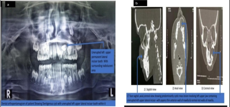
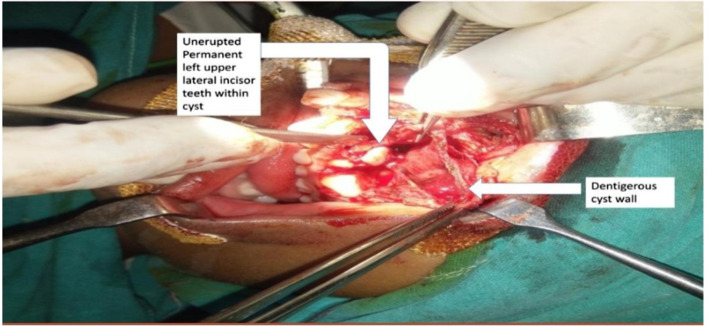
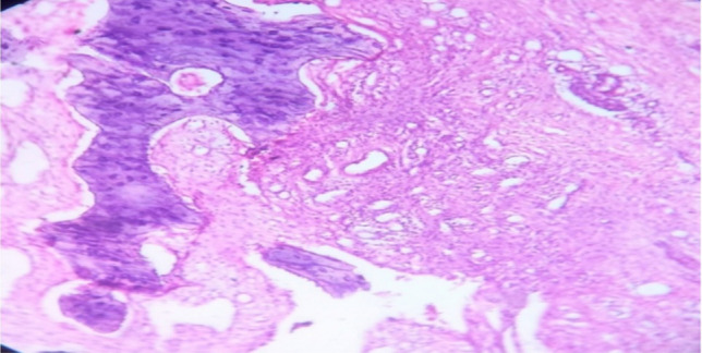
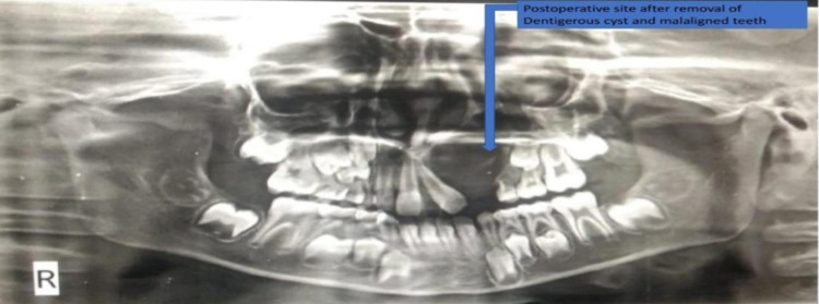
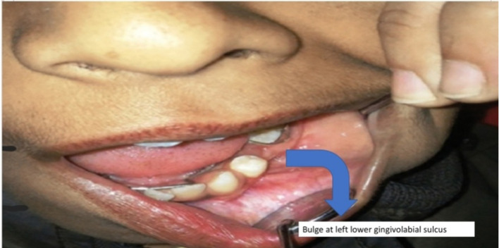
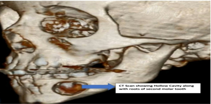
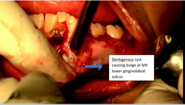
Dentigerous cysts involves erupted or developing teeth in decreasing order of frequency as mandibular third molars, the maxillary canines, the maxillary third molars with rare involvement of the central incisors & are usually asymptomatic but becomes symptomatic with symptoms such as swelling, mild sensitivity, tooth mobility and displacement when it reaches size > 2 cm in diameter. Dentigerous cysts are seen most commonly in 20-30 years age group with relative low frequency in children with proportion of 4-9% of these cysts occur in the first 10 years after birth. Hospital based study conducted in the Department of ENT at Ashwini Rural medical college & Hospital, Solapur in which we have reported a series of three cases of dentigerous cyst in paediatric patients. Dentigerous Cysts in our study were associated with maxillary permanent lateral incisor, mandibular permanent premolar, Mandibular second molar tooth in pediatric age group. Provisional diagnosis of Dentigerous cyst based on clinical, radiological & biochemical study but Histopathological examination confirms the diagnosis. Dental orthopentamogram aids in diagnosing and localizing the Dentigerous cyst and ct face in all views helps to see bony effects and complications due to expansion of Dentigerous cyst. Enucleation of the cystic lesion followed by extraction of the offending teeth is a suitable treatment modality for Dentigerous cyst associated with maxilla and mandible giving adequate balance between complete removal of cyst & giving chance for eruption of unerupted teeth.
Keywords: Dentigerous cyst; Enucleation; Lateral incisor; Orthopentamogram.
© Association of Otolaryngologists of India 2023. Springer Nature or its licensor (e.g. a society or other partner) holds exclusive rights to this article under a publishing agreement with the author(s) or other rightsholder(s); author self-archiving of the accepted manuscript version of this article is solely governed by the terms of such publishing agreement and applicable law.
Conflict of interest statement
Conflict of interestAll the authors declare that they have not any conflict of interest.
Figures


References
-
- Hasbini AS, Hadi U, Ghafari J. Endoscopic removal of an ectopic third molar obstructing the osteomeatal complex. Ear NoseThroat J. 2001;80:667–670. - PubMed
LinkOut - more resources
Full Text Sources
Research Materials
Miscellaneous