

Natural history of intraosseous low-grade chondroid lesions of the proximal humerus
- PMID: 37637054
- PMCID: PMC10457155
- DOI: 10.3389/fonc.2023.1200286
Natural history of intraosseous low-grade chondroid lesions of the proximal humerus
Abstract
Introduction: Enchondromas and grade 1 chondrosarcomas are commonly encountered low-grade chondroid tumors in the proximal humerus. While there is a concern for malignant transformation, few studies have evaluated the natural history of these lesions. The purpose of this study is to evaluate the natural history of proximal humerus low-grade chondroid lesions managed both conservatively and surgically, and to define management criteria using clinical and radiographic findings for these low-grade chondroid lesions.
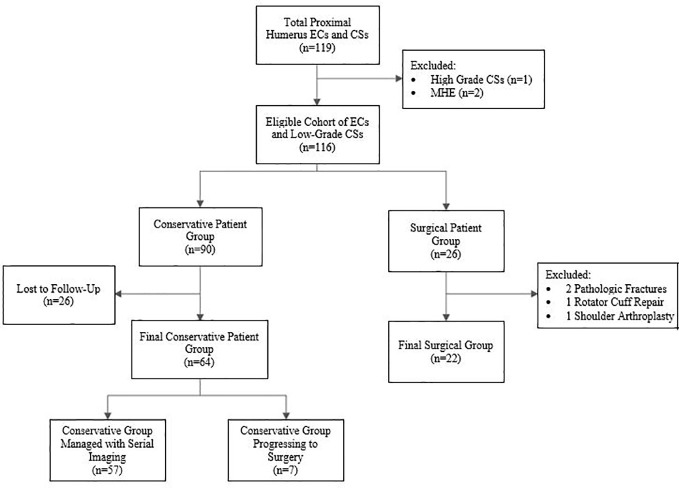
Methods: The patient population included 90 patients intended for conservative treatment and 22 patients proceeding directly to surgery. Data collection was based on a combination of chart review and patient imaging and descriptive statistics were calculated for each group.
Results: No malignant transformations were noted amongst any group. In the conservative treatment group, 7 of 64 (11%) progressed to surgery after an average of 20.3 months of conservative treatment due to persistent pain unexplained by other shoulder pathology. Importantly, 71% experienced continued pain at a mean of 53.1 months post-operatively. The group that went directly to surgery also demonstrated pain in 41% at an average follow-up of 57.3 months.
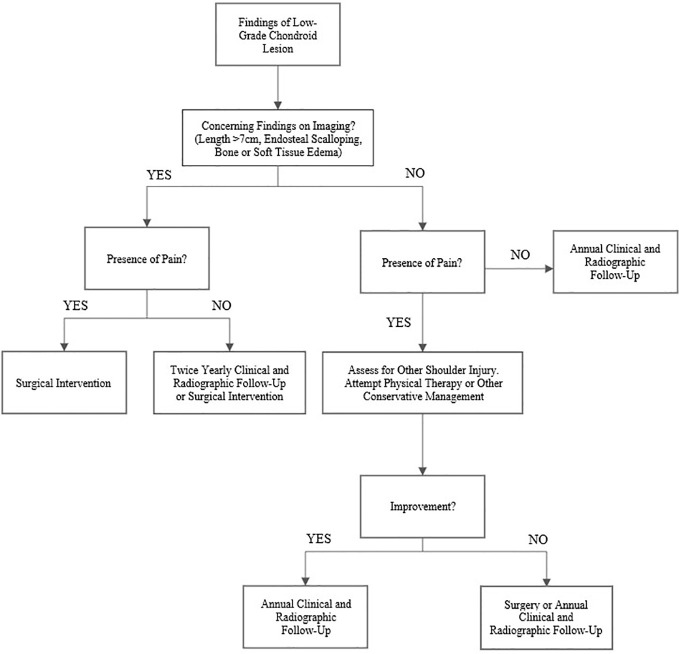
Discussion: Low-grade cartilaginous lesions of the proximal humerus without concerning imaging findings can be managed with conservative treatment and the risk of malignant transformation is very low. Patients with a clear source of their shoulder pain unrelated to their tumor and without concerning characteristics on imaging can be managed with serial annual radiographic imaging. Patients undergoing surgery for these indolent tumors are likely to experience persistent pain even after surgery.
Keywords: cancer/tumors; chondroid; clinical outcomes; diagnostic imaging; shoulder.
Copyright © 2023 LaPrade, Andryk, Christensen, Neilson, Wooldridge, Hackbarth, Bedi and King.
Conflict of interest statement
The authors declare that the research was conducted in the absence of any commercial or financial relationships that could be construed as a potential conflict of interest.
Figures
Similar articles
-
Enchondromas and atypical cartilaginous tumors at the proximal humerus treated with intralesional resection and bone cement filling with or without osteosynthesis: retrospective analysis of 42 cases with 6 years mean follow-up.World J Surg Oncol. 2018 Jul 13;16(1):139. doi: 10.1186/s12957-018-1437-z. World J Surg Oncol. 2018. PMID: 30005680 Free PMC article.
-
Intraosseous atypical chondroid tumor or chondrosarcoma grade 1 in patients with multiple osteochondromas.J Bone Joint Surg Am. 2015 Jan 7;97(1):24-31. doi: 10.2106/JBJS.N.00121. J Bone Joint Surg Am. 2015. PMID: 25568391
-
Radiographic Enchondroma Surveillance: Assessing Clinical Outcomes and Costs Effectiveness.Iowa Orthop J. 2019;39(1):185-193. Iowa Orthop J. 2019. PMID: 31413693 Free PMC article.
-
Chondroid Tumors as Incidental Findings and Differential Diagnosis between Enchondromas and Low-grade Chondrosarcomas.Semin Musculoskelet Radiol. 2019 Feb;23(1):3-18. doi: 10.1055/s-0038-1675550. Epub 2019 Jan 30. Semin Musculoskelet Radiol. 2019. PMID: 30699449 Review.
-
Surgical versus conservative interventions for treating acromioclavicular dislocation of the shoulder in adults.Cochrane Database Syst Rev. 2019 Oct 11;10(10):CD007429. doi: 10.1002/14651858.CD007429.pub3. Cochrane Database Syst Rev. 2019. PMID: 31604007 Free PMC article. Review.
Cited by
-
Microwave ablation combined with curettage for low-grade chondrosarcomas in the extremities: a single-center study.BMC Musculoskelet Disord. 2025 Jul 7;26(1):660. doi: 10.1186/s12891-025-08787-6. BMC Musculoskelet Disord. 2025. PMID: 40624513 Free PMC article.
-
Multiple enchondromas in Ollier's disease: A case report.Radiol Case Rep. 2024 Aug 19;19(11):5033-5037. doi: 10.1016/j.radcr.2024.07.080. eCollection 2024 Nov. Radiol Case Rep. 2024. PMID: 39253051 Free PMC article.
-
A Rare Case of Low-Grade Bilateral Proximal Humerus Chondrosarcomas Managed With Staged Curettage and Cementation.Cureus. 2025 Mar 19;17(3):e80854. doi: 10.7759/cureus.80854. eCollection 2025 Mar. Cureus. 2025. PMID: 40255759 Free PMC article.
References
-
- Brien EW, Mirra JM, Kerr R. Benign and Malignant cartilage tumors of bone and joint: their anatomic and theoretical basis with an emphasis on radiology, pathology and clinical biology. I. The intramedullary cartilage tumors. Skeletal Radiol (1997) 26(6):325–53. doi: 10.1007/s002560050246 - DOI - PubMed
-
- Chang CY, Garner HW, Ahlawat S, Amini B, Bucknor MD, Flug JA, et al. . Society of Skeletal Radiology- white paper. Guidelines for the diagnostic management of incidental solitary bone lesions on CT and MRI in adults: bone reporting and data system (Bone-RADS). Skeletal Radiol (2022) 51(9):1743–64. doi: 10.1007/s00256-022-04022-8 - DOI - PMC - PubMed
-
- Herget GW, Strohm P, Rottenburger C, Kontny U, Krauß T, Bohm J, et al. . Insights into Enchondroma, Enchondromatosis and the risk of secondary Chondrosarcoma. Review of the literature with an emphasis on the clinical behaviour, radiology, Malignant transformation and the follow up. Neoplasma (2014) 61(4):365–78. doi: 10.4149/neo_2014_046 - DOI - PubMed
LinkOut - more resources
Full Text Sources