. 2023 Aug 10:14:1118550.
doi: 10.3389/fphar.2023.1118550. eCollection 2023.

Protective effects of Scutellariae Radix Carbonisata-derived carbon dots on blood-heat and hemorrhage rats

Affiliations

Protective effects of Scutellariae Radix Carbonisata-derived carbon dots on blood-heat and hemorrhage rats

Meiling Zhang et al. Front Pharmacol. .

Abstract

As the charcoal processing product of Scutellariae Radix (SR), SR Carbonisata (SRC) has been clinically used as a cooling blood and hemostatic agent for thousands of years. However, the underlying active ingredients and mechanism of SRC still remained unspecified. In this study, SRC derived carbon dots (SRC-CDs) were extracted and purified from the aqueous solution of SRC, followed by physicochemical property assessment by series of technologies. The cooling blood and hemostatic effects of SRC-CDs were further evaluated via a blood-heat and hemorrhage (BHH) rat model. Results showed that the diameters of obtained fluorescent SRC-CDs ranged from 5.0 nm to 10.0 nm and possessed functional group-rich surfaces. Additionally, the as-prepared SRC-CDs showed remarkable cooling blood and hemostasis effects in BHH model, mainly manifested by significant improvement of elevated rectal temperature, inflammatory cytokines (TNF-α, IL-6, and IL-1β) levels, as well as protein expressions of myD88 and NF-κB p65, abnormal coagulation parameters (elevated APTT and FIB), hemogram parameters (RBC, HGB, and HCT), and histopathological changes in lung and gastric tissues. This study, for the first time, demonstrated that SRC-CDs were the cooling blood and hemostatic active components of SRC, which could inhibit the release of inflammatory cytokines by regulating myD88/NF-κB signaling pathway, and activating the fibrin system and endogenous coagulation pathway. These results not only provide a new perspective for the study of active ingredients of carbonized herbs represented by SRC, but also lay an experimental foundation for the development of next-generation nanomedicines.

Keywords: Scutellariae Radix Carbonisata-derived carbon dots (SRC-CDs); active ingredients; anti-inflammation; cooling blood and hemostatic effects; hemostasis.


Conflict of interest statement

Author TH was employed by the company Merck & Co., Inc. The remaining authors declare that the research was conducted in the absence of any commercial or financial relationships that could be construed as a potential conflict of interest.

Figures

FIGURE 1
FIGURE 1
(A) Flow diagram of preparing process of Scutellariae Radix Carbonisata—derived carbon dots (SRC-CDs) and the solution outside the dialysis membrane of SRC (SRC-OD). (B) HPLC fingerprint of SRC and SRC-CDs. (C) Tail bleeding time of mice (n = 6 per group) treated with normal saline (NS), Yunnan Baiyao (positive), Scutellariae Radix (SR), SRC, SRC-CDs and SRC-OD. Data are represented as mean ± SD. *p < 0.05 vs. NS group or SR group and **p < 0.01 vs. NS group.
FIGURE 2
FIGURE 2
The morphology characterization of Scutellariae Radix Carbonisata—derived carbon dots (SRC-CDs). (A,B) High-resolution transmission electron microscopy (HRTEM) images. Inset: magnification figure. (C) Histogram depicting particle size distribution. (D) Line profiles of the corresponding HRTEM images and (E) X-ray diffraction pattern (XRD) of SRC-CDs.
FIGURE 3
FIGURE 3
The optical characterization of Scutellariae Radix Carbonisata—derived carbon dots (SRC-CDs). (A) Ultraviolet-visible spectrum (UV-Vis), (B) fluorescence spectrum and (C) Fourier transform infrared spectrum (FTIR).
FIGURE 4
FIGURE 4
The X-ray photoelectron spectroscopy (XPS) spectra of Scutellariae Radix Carbonisata—derived carbon dots (SRC-CDs). (A) Full survey spectrum. (B) C 1s spectrum, (C) O 1s spectrum and (D) N 1s spectrum.
FIGURE 5
FIGURE 5
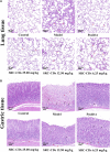
Histopathology of (A) lung tissues and (B) gastric tissues (×100). Sprague Dawley rats were assigned into six groups (n = 10): control (NS), model, positive and Scutellariae Radix Carbonisata—derived carbon dots (SRC-CDs) at high- (25.00 mg/kg), medium- (12.50 mg/kg) and low (6.25 mg/kg) doses groups. The arrows represents local bleeding point, while “△(red)” represents inflammatory cells infiltration.
FIGURE 6
FIGURE 6
Effects of Scutellariae Radix Carbonisata—derived carbon dots (SRC-CDs) on coagulation parameters. (A) Prothrombin time (PT), (B) activated partial thromboplastin time (APTT), (C) thrombin (TT) and (D) fibrinogen (FIB). Sprague Dawley rats were assigned into six groups (n = 10): control, model, positive and SRC-CDs at high- (25.00 mg/kg), medium- (12.50 mg/kg) and low (6.25 mg/kg) doses. *p < 0.05 and **p < 0.01 vs. model group or control group.
FIGURE 7
FIGURE 7
Effects of Scutellariae Radix Carbonisata—derived carbon dots (SRC-CDs) on the levels of inflammatory cytokines. The levels of (A) TNF-α in plasma, (B) TNF-α, (D) IL-1β and (F) IL-6 in lung tissues, (C) TNF-α, (E) IL-1β and (G) IL-6 in gastric tissues. Sprague Dawley rats were assigned into six groups (n = 10): control, model, positive and SRC-CDs at high- (25.00 mg/kg), medium- (12.50 mg/kg) and low (6.25 mg/kg) doses. **p < 0.01 and *p < 0.05 vs. control group or model group.
FIGURE 8
FIGURE 8
Effects of Scutellariae Radix Carbonisata—derived carbon dots (SRC-CDs) on the expression of NF-κB p65 and myD88 proteins in both (A) lung and (B) gastric tissues. (A), (a) and (B), (a) Representative immunoblot analysis and densitometry. (A), (b) and (B), (b) Quantification of NF-κB p 65 protein and myD88 protein. Sprague Dawley rats were assigned into five groups (n = 10): control, model and SRC-CDs at high- (25.00 mg/kg), medium- (12.50 mg/kg) and low (6.25 mg/kg) doses. *p < 0.05 and **p < 0.01 vs. control group or model group.

References

    1. Cao J., Xu X., Jin B., Xiao G. (2012). Functional groups evolvement and charcoal formation during lignin pyrolysis/carbonization. J. Southeast Univ. Sci. Ed. 42, 83–87. 10.3969/j.issn.1001-0505.2012.01.016 - DOI
    1. Chellasamy G., Arumugasamy S. K., Govindaraju S., Yun K. (2021). Green synthesized carbon quantum dots from maple tree leaves for biosensing of Cesium and electrocatalytic oxidation of glycerol. Chemosphere 287 (1), 131915. 10.1016/j.chemosphere.2021.131915 - DOI - PubMed
    1. Cheng J., Zhang M., Sun Z., Lu F., Xiong W., Luo J., et al. (2019a). Hemostatic and hepatoprotective bioactivity of Junci Medulla carbonisata-derived carbon dots. Nanomedicine (Lond) 14 (4), 431–446. 10.2217/nnm-2018-0285 - DOI - PubMed
    1. Cheng P., Xue X. Y., Su J. B., Lu M. M., Wang S. M., Meng J. (2019b). (1)H NMR-based metabonomic revealed protective effect of Moutan Cortex charcoal on blood-heat and hemorrhage rats. J. Pharm. Biomed. Anal. 169, 151–158. 10.1016/j.jpba.2019.02.044 - DOI - PubMed
    1. Fu X., Fu X., Li W. Y., Chen Y. P., Cai Z. X. (2019). Ovalbumin as a precursor for green synthesis of highly fluorescent carbon dots for cell imaging. J. Biomed. Nanotechnol. 15 (6), 1232–1240. 10.1166/jbn.2019.2766 - DOI - PubMed