Review
. 2023 Aug 11:14:1229885.
doi: 10.3389/fimmu.2023.1229885. eCollection 2023.

Lipocalin 2 receptors: facts, fictions, and myths

Affiliations
Review

Lipocalin 2 receptors: facts, fictions, and myths

Sarah K Schröder et al. Front Immunol. .

Abstract

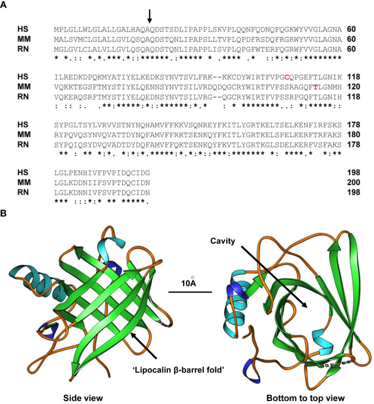
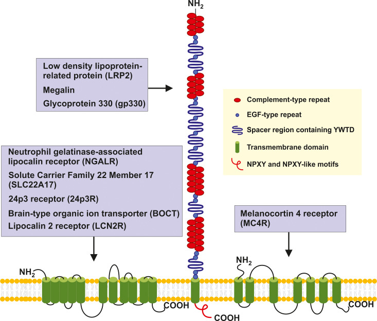
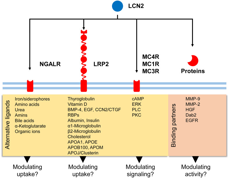
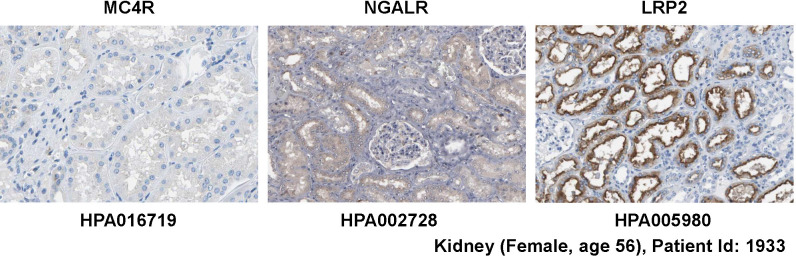
The human 25-kDa Lipocalin 2 (LCN2) was first identified and purified as a protein that in part is associated with gelatinase from neutrophils. This protein shows a high degree of sequence similarity with the deduced sequences of rat α2-microglobulin-related protein and the mouse protein 24p3. Based on its typical lipocalin fold, which consists of an eight-stranded, anti-parallel, symmetrical β-barrel fold structure it was initially thought that LCN2 is a circulating protein functioning as a transporter of small lipophilic molecules. However, studies in Lcn2 null mice have shown that LCN2 has bacteriostatic properties and plays a key role in innate immunity by sequestering bacterial iron siderophores. Numerous reports have further shown that LCN2 is involved in the control of cell differentiation, energy expenditure, cell death, chemotaxis, cell migration, and many other biological processes. In addition, important roles for LCN2 in health and disease have been identified in Lcn2 null mice and multiple molecular pathways required for regulation of Lcn2 expression have been identified. Nevertheless, although six putative receptors for LCN2 have been proposed, there is a fundamental lack in understanding of how these cell-surface receptors transmit and amplify LCN2 to the cell. In the present review we summarize the current knowledge on LCN2 receptors and discuss inconsistencies, misinterpretations and false assumptions in the understanding of these potential LCN2 receptors.

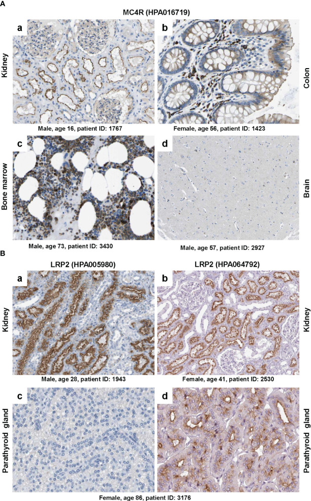
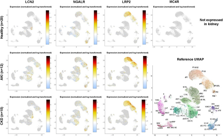
Keywords: LRP2; MC4R; Megalin; NGALR; SLC22A17; inflammation; iron.


Conflict of interest statement

The authors declare that the research was conducted in the absence of any commercial or financial relationships that could be construed as a potential conflict of interest.

Figures

Figure 1
Figure 1
Structure of LCN2. (A) Alignment of human (HS), mouse (MM) and rat (RN) LCN2 proteins. The side of cleavage of the secretory signal peptide is marked by a black arrow. In addition, the tyrosine in mouse LCN2 (T115) shown to be a PKCδ phosphorylation site and the cysteine in human LCN2 (C107) shown to form the disulfide bridge with human MMP-9 is marked in red letters. An asterisk (*) indicates fully conserved residues, a colon (:) conservation between groups of strongly similar properties, and a period (.) conservation between groups of weakly similar properties. Gaps in the aligned sequences are indicated by dashes. (B) Side (left) and bottom-to-top (right) views of human LCN2 showing the typical eight-stranded, anti-parallel, symmetrical β-barrel fold that is characteristic of members of the lipocalin family. The formed cavity is supposed to permit binding of a broad array of bulky ligands. The depicted structure of the apo-form of human LCN2 determined by X-ray diffraction resolved at resolution of 2Å was generated using the Ribbons XP software (version 3.0) and structure coordinates deposited in the RCSB Protein Data Bank (4) [http://www.rcsb.org] under accession no. 3BX8. A size marker (10Å) is given.
Figure 2
Figure 2
Structure and potential signaling pathways targeted by the putative LCN2 receptors. The three putative receptors for LCN2 are structurally different. NGALR also known as SLC22A17, 24p3, BOCT or LCN2R is a multipass 60-kDa integral membrane protein predicted to contain 11-12 transmembrane helices that are linked by extracellular and intercellular spacers of variable sizes (13, 29, 30). LRP2/megalin/gp330 is a ~4,600 amino-acid type 1 transmembrane receptor of the LDL receptor gene family characterized by extracellular domains containing four cysteine-rich clusters of complement-type repeats (i.e., the low-density lipoprotein-receptor type A repeats) that mediate ligand binding that are separated and followed by 17 epidermal growth factor type repeats and eight spacer regions that contain YWTD repeats. These are termed β-propellers that are required for pH-dependent release of bound ligands in endosomes (31). The single transmembrane of LRP2 encompassing 20 amino acids is followed by a 213 amino acid cytoplasmic tail, which contains two NPXY sequences and one NPXY-like sequence in addition to several Src-homology 3 (SH3) and one Src-homolog-2 (SH2) region sites (32). MC4R is a 332 amino acid G protein-coupled receptor (GPCR) with seven transmembrane helixes connected by alternating extracellular and intercellular loops. The recent structure of the human MC4R-GS signaling complex bound to the agonist setmalanotide determined by electron microscopy demonstrated that the seven transmembrane-spanning helixes from a bundle that forms a cavity at the cytoplasmic side to accommodate the heterotrimeric Gs protein (33, 34). For simplicity the seven helixes are drawn as stand-alone transmembrane domains.
Figure 3
Figure 3
Potential signaling routes for LCN2. LCN2 has affinity for five putative receptors (NGALR, LRP2, MC4R, MC1R, and MC3R) and additional binding partners (e.g., MMP-9, MMP-2, HGF). Binding of LCN2 to these biomolecules can modulate activity of respective proteins/receptors or modulate signaling cascades triggered. In addition to these receptors, LCN2 was recently found to bind to LRP6 in mouse embryonic fibroblasts (49).
Figure 4
Figure 4
Expression of potential LCN2 receptors in kidney. Normal kidney tissue sections were stained with antibodies specific for MC4R, NGALR, and LRP2 showing that the kidney lacks MC4R expression. All images were taken from the Human Protein Atlas (91) database [https://www.proteinatlas.org/ 92]. The images can be found at: https://www.proteinatlas.org/ENSG00000166603-MC4R/tissue/kidney#img (left), https://www.proteinatlas.org/ENSG00000092096-SLC22A17/tissue/kidney#img (middle), and https://www.proteinatlas.org/ENSG00000081479-LRP2/tissue/kidney#img (right).
Figure 5
Figure 5
Immunohistochemical staining of MC4R and LRP2/megalin. (A) Tissue sections from (a) kidney, (b) colon, (c) bone marrow and (d) brain were stained with a validated antibody (HPA016719) specific for MC4R. Please note that the antibody stained tubular cells in the kidney, hematopoietic cells in the bone marrow, glandular cells in the colon, while only showing low staining in some neuronal cells in the brain. (B) Tissue sections from (a, b) kidney and (c, d) parathyroid gland were stained with two (validated) polyclonal rabbit antibodies (i.e., HPA005980 and HPA064792) directed against human LRP2. Please note that both antibodies stained similar structures in kidney, while the staining was markedly different in two sections of the parathyroid gland that were taken from the same patient. All images were taken from the Human Protein Atlas (91) database [https://www.proteinatlas.org/ 92]. The images depicted in (A) can be found at: (a) https://www.proteinatlas.org/ENSG00000166603-MC4R/tissue/kidney#img, (b) https://www.proteinatlas.org/ENSG00000166603-MC4R/tissue/colon#img, (c), https://www.proteinatlas.org/ENSG00000166603-MC4R/tissue/bone+marrow#img, and (d) https://www.proteinatlas.org/ENSG00000166603-MC4R/tissue/cerebral+cortex#img. The images depicted in (B) can be found at: (a, b) https://www.proteinatlas.org/ENSG00000081479-LRP2/tissue/kidney#img and (c, d) https://www.proteinatlas.org/ENSG00000081479-LRP2/tissue/parathyroid+gland#img.
Figure 6
Figure 6
Expression of LCN2 and its putative receptors in human kidney in health and disease as obtained from single-cell RNA-sequencing. The dataset comprises 20 samples from 18 living donor biopsy participants taken from the Human Cell Atlas, as well as 15 biopsies from patients suffering from chronic kidney disease and 12 biopsies taken from patients suffering from acute kidney injury. For individual abbreviations depicted in the two-dimensional Reference Uniform Manifold Approximation and Projection (UMAP) image, please refer to Supplementary Table 1 . Please note that the cell subsets that are capable to express LCN2 increase during renal disease, while the expression profile of the putative LCN2 receptors NGALR and LRP2 does not change. Moreover, renal expression of LRP2 is more restricted and MC4R is not expressed at all in any kidney cell fraction. The results here are in whole or part based upon data generated by KPMP [https://atlas.kpmp.org]. The respective URL address used for visualizing expression data from LCN2, NGALR, LRP2, and MC4R in healthy kidney, acute kidney injury and chronic kidney disease is: https://atlas.kpmp.org/explorer/dataviz. All data were downloaded on 20 May, 2023.

Similar articles

Cited by

References

    1. Chan YL, Paz V, Wool IG. The primary structure of rat alpha 2µ globulin-related protein. Nucleic Acids Res (1988) 16(23):11368. doi: 10.1093/nar/16.23.1136 - DOI - PMC - PubMed
    1. Hraba-Renevey S, Türler H, Kress M, Salomon C, Weil R. SV40-induced expression of mouse gene 24p3 involves a post-transcriptional mechanism. Oncogene (1989) 4(5):601–8. - PubMed
    1. Kjeldsen L, Johnsen AH, Sengeløv H, Borregaard N. Isolation and primary structure of NGAL, a novel protein associated with human neutrophil gelatinase. J Biol Chem (1993) 268(14):10425–32. doi: 10.1016/S0021-9258(18)82217-7 - DOI - PubMed
    1. RCSB . Protein Data Bank (PDB). Available at: https://www.rcsb.org/ (Accessed 11 May 2023).
    1. Nilsen-Hamilton M, Hamilton RT, Adams GA. Rapid selective stimulation by growth factors of the incorporation by BALB/C 3T3 cells of [35S]methionine into a glycoprotein and five superinducible proteins. Biochem Biophys Res Commun (1982) 108(1):158–66. doi: 10.1016/0006-291x(82)91845-9 - DOI - PubMed

Publication types